iBet uBet web content aggregator. Adding the entire web to your favor.


Link to original content: https://www.rcsb.org/structure/3bht
RCSB PDB - 3BHT: Structure of phosphorylated Thr160 CDK2/cyclin A in complex with the inhibitor meriolin 3

 3BHT

Structure of phosphorylated Thr160 CDK2/cyclin A in complex with the inhibitor meriolin 3


Experimental Data Snapshot

  • Method: X-RAY DIFFRACTION
  • Resolution: 2.00 Å
  • R-Value Free: 0.220 
  • R-Value Work: 0.181 
  • R-Value Observed: 0.183 

wwPDB Validation   3D Report Full Report


Ligand Structure Quality Assessment 


This is version 1.2 of the entry. See complete history


Literature

Meriolins, a new class of cell death inducing kinase inhibitors with enhanced selectivity for cyclin-dependent kinases

Bettayeb, K.Tirado, O.M.Marionneau-Lambot, S.Ferandin, Y.Lozach, O.Morris, J.C.Mateo-Lozano, S.Drueckes, P.Kubbutat, M.H.Liger, F.Marquet, B.Joseph, B.Echalier, A.Endicott, J.A.Notario, V.Meijer, L.

(2007) Cancer Res 67: 8325-8334

  • DOI: https://doi.org/10.1158/0008-5472.CAN-07-1826
  • Primary Citation of Related Structures:  
    3BHT, 3BHU, 3BHV

  • PubMed Abstract: 

    Protein kinases represent promising anticancer drug targets. We describe here the meriolins, a new family of inhibitors of cyclin-dependent kinases (CDK). Meriolins represent a chemical structural hybrid between meridianins and variolins, two families of kinase inhibitors extracted from various marine invertebrates. Variolin B is currently in preclinical evaluation as an antitumor agent. A selectivity study done on 32 kinases showed that, compared with variolin B, meriolins display enhanced specificity toward CDKs, with marked potency on CDK2 and CDK9. The structures of pCDK2/cyclin A/variolin B and pCDK2/cyclin A/meriolin 3 complexes reveal that the two inhibitors bind within the ATP binding site of the kinase, but in different orientations. Meriolins display better antiproliferative and proapoptotic properties in human tumor cell cultures than their parent molecules, meridianins and variolins. Phosphorylation at CDK1, CDK4, and CDK9 sites on, respectively, protein phosphatase 1alpha, retinoblastoma protein, and RNA polymerase II is inhibited in neuroblastoma SH-SY5Y cells exposed to meriolins. Apoptosis triggered by meriolins is accompanied by rapid Mcl-1 down-regulation, cytochrome c release, and activation of caspases. Meriolin 3 potently inhibits tumor growth in two mouse xenograft cancer models, namely, Ewing's sarcoma and LS174T colorectal carcinoma. Meriolins thus constitute a new CDK inhibitory scaffold, with promising antitumor activity, derived from molecules initially isolated from marine organisms.


  • Organizational Affiliation

    Centre National de la Recherche Scientifique, Cell Cycle Group & UPS2682, Station Biologique, Roscoff, Bretagne, France.


Macromolecules
Find similar proteins by:  (by identity cutoff)  |  3D Structure
Entity ID: 1
MoleculeChains Sequence LengthOrganismDetailsImage
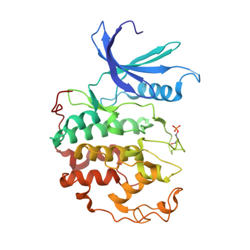
Cell division protein kinase 2
A, C
300Homo sapiensMutation(s): 0 
Gene Names: CDK2
EC: 2.7.11.22
UniProt & NIH Common Fund Data Resources
Find proteins for P24941 (Homo sapiens)
Explore P24941 
Go to UniProtKB:  P24941
PHAROS:  P24941
GTEx:  ENSG00000123374 
Entity Groups  
Sequence Clusters30% Identity50% Identity70% Identity90% Identity95% Identity100% Identity
UniProt GroupP24941
Sequence Annotations
Expand
  • Reference Sequence
Find similar proteins by:  (by identity cutoff)  |  3D Structure
Entity ID: 2
MoleculeChains Sequence LengthOrganismDetailsImage
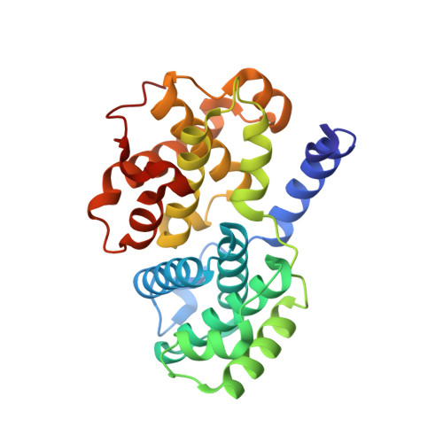
Cyclin-A2
B, D
262Bos taurusMutation(s): 0 
Gene Names: CCNA2CCNA
UniProt
Find proteins for P30274 (Bos taurus)
Explore P30274 
Go to UniProtKB:  P30274
Entity Groups  
Sequence Clusters30% Identity50% Identity70% Identity90% Identity95% Identity100% Identity
UniProt GroupP30274
Sequence Annotations
Expand
  • Reference Sequence
Small Molecules
Ligands 3 Unique
IDChains Name / Formula / InChI Key2D Diagram3D Interactions
MFR
Query on MFR

Download Ideal Coordinates CCD File 
E [auth A],
I [auth C]
4-(4-methoxy-1H-pyrrolo[2,3-b]pyridin-3-yl)pyrimidin-2-amine
C12 H11 N5 O
GGEWEQJWGGJUHO-UHFFFAOYSA-N
SGM
Query on SGM

Download Ideal Coordinates CCD File 
G [auth B],
H [auth B],
K [auth D]
MONOTHIOGLYCEROL
C3 H8 O2 S
PJUIMOJAAPLTRJ-GSVOUGTGSA-N
MG
Query on MG

Download Ideal Coordinates CCD File 
F [auth B],
J [auth D]
MAGNESIUM ION
Mg
JLVVSXFLKOJNIY-UHFFFAOYSA-N
Modified Residues  1 Unique
IDChains TypeFormula2D DiagramParent
TPO
Query on TPO
A, C
L-PEPTIDE LINKINGC4 H10 N O6 PTHR
Binding Affinity Annotations 
IDSourceBinding Affinity
MFR BindingDB:  3BHT IC50: 11 (nM) from 1 assay(s)
Experimental Data & Validation

Experimental Data

  • Method: X-RAY DIFFRACTION
  • Resolution: 2.00 Å
  • R-Value Free: 0.220 
  • R-Value Work: 0.181 
  • R-Value Observed: 0.183 
  • Space Group: P 21 21 21
Unit Cell:
Length ( Å )Angle ( ˚ )
a = 74.128α = 90
b = 133.77β = 90
c = 147.831γ = 90
Software Package:
Software NamePurpose
MOSFLMdata reduction
SCALAdata scaling
REFMACrefinement
PDB_EXTRACTdata extraction
ADSCdata collection
MOLREPphasing

Structure Validation

View Full Validation Report


Ligand Structure Quality Assessment 


Entry History 

Revision History  (Full details and data files)

  • Version 1.0: 2008-02-12
    Type: Initial release
  • Version 1.1: 2011-07-13
    Changes: Advisory, Version format compliance
  • Version 1.2: 2024-10-30
    Changes: Data collection, Database references, Derived calculations, Structure summary