iBet uBet web content aggregator. Adding the entire web to your favor.


Link to original content: https://www.rcsb.org/structure/2M5B
RCSB PDB - 2M5B: The NMR structure of the BID-BAK complex

 2M5B

The NMR structure of the BID-BAK complex


Experimental Data Snapshot

  • Method: SOLUTION NMR
  • Conformers Calculated: 100 
  • Conformers Submitted: 20 
  • Selection Criteria: structures with the lowest energy 

wwPDB Validation   3D Report Full Report


This is version 2.1 of the entry. See complete history


Literature

BID-induced structural changes in BAK promote apoptosis.

Moldoveanu, T.Grace, C.R.Llambi, F.Nourse, A.Fitzgerald, P.Gehring, K.Kriwacki, R.W.Green, D.R.

(2013) Nat Struct Mol Biol 20: 589-597

  • DOI: https://doi.org/10.1038/nsmb.2563
  • Primary Citation of Related Structures:  
    2M5B

  • PubMed Abstract: 

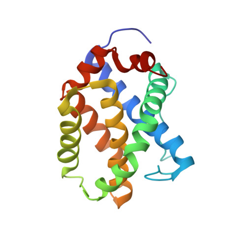
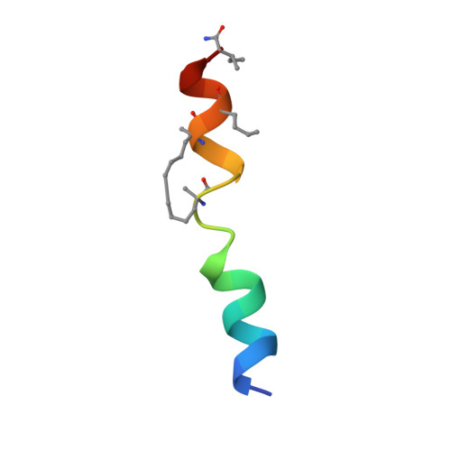
    The BCL-2-family protein BAK is responsible for mitochondrial outer-membrane permeabilization (MOMP), which leads to apoptosis. The BCL-2 homology 3 (BH3)-only protein BID activates BAK to perform this function. We report the NMR solution structure of the human BID BH3-BAK complex, which identified the activation site at the canonical BH3-binding groove of BAK. Mutating the BAK BH1 in the groove prevented activation and MOMP but not the binding of BID. BAK BH3 mutations allowed BID binding and activation but blunted function by blocking BAK oligomerization. BAK activation follows a 'hit-and-run' mechanism whereby BID dissociates from the trigger site, which allows BAK oligomerization at an overlapping interface. In contrast, the BH3-only proteins NOXA and BAD are predicted to clash with the trigger site and are not activators of BAK. These findings provide insights into the early stages of BAK activation.


  • Organizational Affiliation

    Department of Immunology, St. Jude Children's Research Hospital, Memphis, Tennessee, USA.


Macromolecules
Find similar proteins by:  (by identity cutoff)  |  3D Structure
Entity ID: 1
MoleculeChains Sequence LengthOrganismDetailsImage
Bcl-2 homologous antagonist/killer169Homo sapiensMutation(s): 0 
Gene Names: BAKBAK1BCL2L7CDN1
UniProt & NIH Common Fund Data Resources
Find proteins for Q16611 (Homo sapiens)
Explore Q16611 
Go to UniProtKB:  Q16611
PHAROS:  Q16611
GTEx:  ENSG00000030110 
Entity Groups  
Sequence Clusters30% Identity50% Identity70% Identity90% Identity95% Identity100% Identity
UniProt GroupQ16611
Sequence Annotations
Expand
  • Reference Sequence

Find similar proteins by:  Sequence   |   3D Structure  

Entity ID: 2
MoleculeChains Sequence LengthOrganismDetailsImage
human_BID_BH3_SAHB23synthetic constructMutation(s): 0 
UniProt & NIH Common Fund Data Resources
Find proteins for P55957 (Homo sapiens)
Explore P55957 
Go to UniProtKB:  P55957
PHAROS:  P55957
GTEx:  ENSG00000015475 
Entity Groups  
Sequence Clusters30% Identity50% Identity70% Identity90% Identity95% Identity100% Identity
UniProt GroupP55957
Sequence Annotations
Expand
  • Reference Sequence
Small Molecules
Modified Residues  2 Unique
IDChains TypeFormula2D DiagramParent
MK8
Query on MK8
B
L-PEPTIDE LINKINGC7 H15 N O2LEU
NLE
Query on NLE
B
L-PEPTIDE LINKINGC6 H13 N O2LEU
Experimental Data & Validation

Experimental Data

  • Method: SOLUTION NMR
  • Conformers Calculated: 100 
  • Conformers Submitted: 20 
  • Selection Criteria: structures with the lowest energy 

Structure Validation

View Full Validation Report


Entry History 

Deposition Data

Revision History  (Full details and data files)

  • Version 1.0: 2013-04-17
    Type: Initial release
  • Version 1.1: 2013-05-08
    Changes: Database references
  • Version 1.2: 2013-05-22
    Changes: Database references
  • Version 1.3: 2023-06-14
    Changes: Data collection, Database references, Derived calculations, Other
  • Version 2.0: 2023-11-15
    Changes: Atomic model, Data collection
  • Version 2.1: 2024-10-09
    Changes: Database references, Structure summary