iBet uBet web content aggregator. Adding the entire web to your favor.


Link to original content: https://www.rcsb.org/structure/1rco
RCSB PDB - 1RCO: SPINACH RUBISCO IN COMPLEX WITH THE INHIBITOR D-XYLULOSE-2,2-DIOL-1,5-BISPHOSPHATE

 1RCO

SPINACH RUBISCO IN COMPLEX WITH THE INHIBITOR D-XYLULOSE-2,2-DIOL-1,5-BISPHOSPHATE


Experimental Data Snapshot

  • Method: X-RAY DIFFRACTION
  • Resolution: 2.30 Å
  • R-Value Free: 0.246 
  • R-Value Work: 0.239 
  • R-Value Observed: 0.239 

Starting Model: experimental
View more details

wwPDB Validation   3D Report Full Report


Ligand Structure Quality Assessment 


This is version 1.4 of the entry. See complete history


Literature

A common structural basis for the inhibition of ribulose 1,5-bisphosphate carboxylase by 4-carboxyarabinitol 1,5-bisphosphate and xylulose 1,5-bisphosphate.

Taylor, T.C.Fothergill, M.D.Andersson, I.

(1996) J Biol Chem 271: 32894-32899

  • DOI: https://doi.org/10.1074/jbc.271.51.32894
  • Primary Citation of Related Structures:  
    1RBO, 1RCO

  • PubMed Abstract: 

    Ribulose 1,5-bisphosphate carboxylase/oxygenase (Rubisco) catalyzes the carboxylation of ribulose 1,5-bisphosphate. The reaction catalyzed by Rubisco involves several steps, some of which can occur as partial reactions, forming intermediates that can be isolated. Analogues of these intermediates are potent inhibitors of the enzyme. We have studied the interactions with the enzyme of two inhibitors, xylulose 1,5-bisphosphate and 4-carboxyarabinitol 1,5-bisphosphate, by x-ray crystallography. Crystals of the complexes were formed by cocrystallization under activating conditions. In addition, 4-carboxyarabinitol 1,5-bisphosphate was soaked into preformed activated crystals of the enzyme. The result of these experiments was the release of the activating CO2 molecule as well as the metal ion from the active site when the inhibitors bound to the enzyme. Comparison with the structure of an activated complex of the enzyme indicates that the structural basis for the release of the activator groups is a distortion of the metal binding site due to the different geometry of the C-3 hydroxyl of the inhibitors. Both inhibitors induce closure of active site loops despite the inactivated state of the enzyme. Xylulose 1,5-bisphosphate binds in a hydrated form at the active site.


  • Organizational Affiliation

    Department of Molecular Biology, Swedish University of Agricultural Sciences, P. O. Box 590, S-751 24 Uppsala, Sweden.


Macromolecules
Find similar proteins by:  (by identity cutoff)  |  3D Structure
Entity ID: 1
MoleculeChains Sequence LengthOrganismDetailsImage
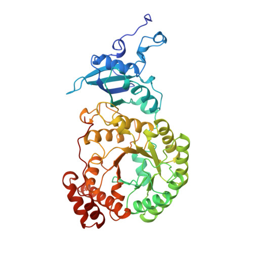
RIBULOSE BISPHOSPHATE CARBOXYLASE/OXYGENASE475Spinacia oleraceaMutation(s): 0 
EC: 4.1.1.39
UniProt
Find proteins for P00875 (Spinacia oleracea)
Explore P00875 
Go to UniProtKB:  P00875
Entity Groups  
Sequence Clusters30% Identity50% Identity70% Identity90% Identity95% Identity100% Identity
UniProt GroupP00875
Sequence Annotations
Expand
  • Reference Sequence
Find similar proteins by:  (by identity cutoff)  |  3D Structure
Entity ID: 2
MoleculeChains Sequence LengthOrganismDetailsImage
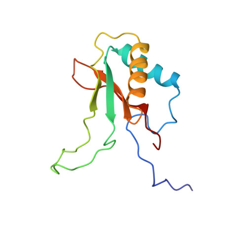
RIBULOSE BISPHOSPHATE CARBOXYLASE/OXYGENASE123Spinacia oleraceaMutation(s): 0 
EC: 4.1.1.39
UniProt
Find proteins for P00870 (Spinacia oleracea)
Explore P00870 
Go to UniProtKB:  P00870
Entity Groups  
Sequence Clusters30% Identity50% Identity70% Identity90% Identity95% Identity100% Identity
UniProt GroupP00870
Sequence Annotations
Expand
  • Reference Sequence
Experimental Data & Validation

Experimental Data

  • Method: X-RAY DIFFRACTION
  • Resolution: 2.30 Å
  • R-Value Free: 0.246 
  • R-Value Work: 0.239 
  • R-Value Observed: 0.239 
  • Space Group: P 21 21 2
Unit Cell:
Length ( Å )Angle ( ˚ )
a = 220.6α = 90
b = 221.7β = 90
c = 115.3γ = 90
Software Package:
Software NamePurpose
DENZOdata reduction
SCALEPACKdata scaling
X-PLORmodel building
X-PLORrefinement
X-PLORphasing

Structure Validation

View Full Validation Report


Ligand Structure Quality Assessment 


Entry History 

Deposition Data

Revision History  (Full details and data files)

  • Version 1.0: 1997-03-12
    Type: Initial release
  • Version 1.1: 2008-03-24
    Changes: Version format compliance
  • Version 1.2: 2011-07-13
    Changes: Version format compliance
  • Version 1.3: 2023-08-09
    Changes: Database references, Derived calculations, Refinement description
  • Version 1.4: 2024-10-30
    Changes: Data collection, Structure summary