iBet uBet web content aggregator. Adding the entire web to your favor.


Link to original content: https://www.rcsb.org/structure/1HFS
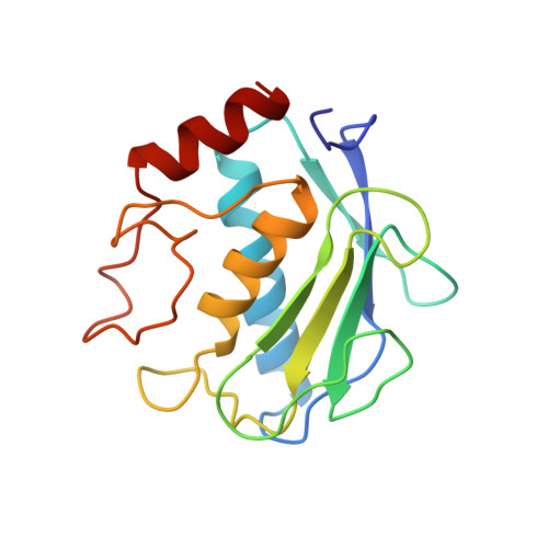
RCSB PDB - 1HFS: CRYSTAL STRUCTURE OF THE CATALYTIC DOMAIN OF HUMAN FIBROBLAST STROMELYSIN-1 INHIBITED WITH THE N-CARBOXY-ALKYL INHIBITOR L-764,004

 1HFS

CRYSTAL STRUCTURE OF THE CATALYTIC DOMAIN OF HUMAN FIBROBLAST STROMELYSIN-1 INHIBITED WITH THE N-CARBOXY-ALKYL INHIBITOR L-764,004


Experimental Data Snapshot

  • Method: X-RAY DIFFRACTION
  • Resolution: 1.70 Å
  • R-Value Free: 0.217 
  • R-Value Work: 0.189 
  • R-Value Observed: 0.189 

wwPDB Validation   3D Report Full Report


Ligand Structure Quality Assessment 


This is version 2.0 of the entry. See complete history


Literature

Inhibition of stromelysin-1 (MMP-3) by P1'-biphenylylethyl carboxyalkyl dipeptides.

Esser, C.K.Bugianesi, R.L.Caldwell, C.G.Chapman, K.T.Durette, P.L.Girotra, N.N.Kopka, I.E.Lanza, T.J.Levorse, D.A.MacCoss, M.Owens, K.A.Ponpipom, M.M.Simeone, J.P.Harrison, R.K.Niedzwiecki, L.Becker, J.W.Marcy, A.I.Axel, M.G.Christen, A.J.McDonnell, J.Moore, V.L.Olszewski, J.M.Saphos, C.Visco, D.M.Shen, F.Colletti, A.Krieter, P.A.Hagmann, W.K.

(1997) J Med Chem 40: 1026-1040

  • DOI: https://doi.org/10.1021/jm960465t
  • Primary Citation of Related Structures:  
    1HFS

  • PubMed Abstract: 

    Carboxyalkyl peptides containing a biphenylylethyl group at the P1' position were found to be potent inhibitors of stromelysin-1 (MMP-3) and gelatinase A (MMP-2), in the range of 10-50 nM, but poor inhibitors of collagenase (MMP-1). Combination of a biphenylylethyl moiety at P1', a tert-butyl group at P2', and a methyl group at P3' produced orally bioavailable inhibitors as measured by an in vivo model of MMP-3 degradation of radiolabeled transferrin in the mouse pleural cavity. The X-ray structure of a complex of a P1-biphenyl inhibitor and the catalytic domain of MMP-3 is described. Inhibitors that contained halogenated biphenylylethyl residues at P1' proved to be superior in terms of enzyme potency and oral activity with 2(R)-[2-(4'-fluoro-4-biphenylyl)ethyl]-4(S)-n-butyl-1,5-pentane dioic acid 1-(alpha(S)-tert-butylglycine methylamide) amide (L-758,354, 26) having a Ki of 10 nM against MMP-3 and an ED50 of 11 mg/kg po in the mouse pleural cavity assay. This compound was evaluated in acute (MMP-3 and IL-1 beta injection in the rabbit) and chronic (rat adjuvant-induced arthritis and mouse collagen-induced arthritis) models of cartilage destruction but showed activity only in the MMP-3 injection model (ED50 = 6 mg/kg iv).


  • Organizational Affiliation

    Department of Medicinal Chemistry, Merck Research Laboratories, Rahway, New Jersey 07065-0900, USA.


Macromolecules
Find similar proteins by:  (by identity cutoff)  |  3D Structure
Entity ID: 1
MoleculeChains Sequence LengthOrganismDetailsImage
STROMELYSIN-1160Homo sapiensMutation(s): 0 
EC: 3.4.24.17
UniProt & NIH Common Fund Data Resources
Find proteins for P08254 (Homo sapiens)
Explore P08254 
Go to UniProtKB:  P08254
PHAROS:  P08254
GTEx:  ENSG00000149968 
Entity Groups  
Sequence Clusters30% Identity50% Identity70% Identity90% Identity95% Identity100% Identity
UniProt GroupP08254
Sequence Annotations
Expand
  • Reference Sequence
Small Molecules
Ligands 3 Unique
IDChains Name / Formula / InChI Key2D Diagram3D Interactions
L04
Query on L04

Download Ideal Coordinates CCD File 
G [auth A]6-(4'-FLUORO-BIPHENYL-4-YL)-4-(3-METHYL-1-PHENYLCARBAMOYL-BUTYLCARBAMOYL)-2-[4-(1-OXO-1,3-DIHYDRO-ISOINDOL-2-YL)-BUTYL]-HEXANOIC ACID
C43 H48 F N3 O5
WSHMSPGZDPZGRV-GZYXPWJKSA-N
ZN
Query on ZN

Download Ideal Coordinates CCD File 
B [auth A],
C [auth A]
ZINC ION
Zn
PTFCDOFLOPIGGS-UHFFFAOYSA-N
CA
Query on CA

Download Ideal Coordinates CCD File 
D [auth A],
E [auth A],
F [auth A]
CALCIUM ION
Ca
BHPQYMZQTOCNFJ-UHFFFAOYSA-N
Binding Affinity Annotations 
IDSourceBinding Affinity
L04 BindingDB:  1HFS Ki: 2 (nM) from 1 assay(s)
PDBBind:  1HFS Ki: 2 (nM) from 1 assay(s)
Experimental Data & Validation

Experimental Data

  • Method: X-RAY DIFFRACTION
  • Resolution: 1.70 Å
  • R-Value Free: 0.217 
  • R-Value Work: 0.189 
  • R-Value Observed: 0.189 
  • Space Group: C 2 2 21
Unit Cell:
Length ( Å )Angle ( ˚ )
a = 84.14α = 90
b = 85.35β = 90
c = 55.26γ = 90
Software Package:
Software NamePurpose
X-PLORmodel building
X-PLORrefinement
R-AXISdata reduction
X-PLORphasing

Structure Validation

View Full Validation Report


Ligand Structure Quality Assessment 


Entry History 

Deposition Data

  • Released Date: 1998-02-18 
  • Deposition Author(s): Becker, J.W.

Revision History  (Full details and data files)

  • Version 1.0: 1998-02-18
    Type: Initial release
  • Version 1.1: 2008-03-24
    Changes: Version format compliance
  • Version 1.2: 2011-07-13
    Changes: Version format compliance
  • Version 1.3: 2012-02-22
    Changes: Database references
  • Version 1.4: 2017-11-29
    Changes: Derived calculations, Other
  • Version 2.0: 2024-02-07
    Changes: Atomic model, Data collection, Database references, Derived calculations