iBet uBet web content aggregator. Adding the entire web to your favor.


Link to original content: https://www.ncbi.nlm.nih.gov/pubmed/28812422
Novel Influenza D virus: Epidemiology, pathology, evolution and biological characteristics - PubMed Skip to main page content
U.S. flag

An official website of the United States government

Dot gov

The .gov means it’s official.
Federal government websites often end in .gov or .mil. Before sharing sensitive information, make sure you’re on a federal government site.

Https

The site is secure.
The https:// ensures that you are connecting to the official website and that any information you provide is encrypted and transmitted securely.

Access keys NCBI Homepage MyNCBI Homepage Main Content Main Navigation
Review
. 2017 Nov 17;8(8):1580-1591.
doi: 10.1080/21505594.2017.1365216. Epub 2017 Aug 25.

Novel Influenza D virus: Epidemiology, pathology, evolution and biological characteristics

Affiliations
Review

Novel Influenza D virus: Epidemiology, pathology, evolution and biological characteristics

Shuo Su et al. Virulence. .

Abstract

In 2011, a new virus was isolated from pigs with influenza-like symptoms and subsequently also from cattle, which are the main reservoir of the virus. It is similar to Influenza C virus (ICV), a (predominantly) human pathogen, causing respiratory disease in children. Since the virus is unable to reassort with ICV (and based on several other criteria as discussed in the text) it is now officially named as Influenzavirus D (IDV), a new genus of the Orthomyxoviridae. We summarize the epidemiology, pathology and evolution of IDV and its biological characteristics with emphasis on the only glycoprotein HEF. Based on the limited data available we finally consider whether IDV represent a public health threat.

Keywords: HEF; Influenzavirus D; bovine; cattle; epidemiology; evolution; pathology; zoonosis.


Figures

Figure 1.
Figure 1.
Reservoir host mode diagram of IDV. IDV was initially isolated from pigs and subsequently from bovine. Since the seroprevalence in bovine is much higher than in swine, bovine is considered to be the primary natural reservoir for IDV. IDV can be transmitted between cattle and also between pigs as indicated by the solid line. Antibody specific for IDV were also detected in sera form small ruminants (sheep and goat) and from humans, especially in people with cattle exposure, but no virus was isolated (indicated by dotted line), indicating that IDV may be transmitted from cattle to humans (dotted line).
Figure 2.
Figure 2.
Structure of the 7 influenza D virus genome segments. Influenza D virus particles possess 7 segments of minus-sense, single-stranded RNA. The open reading frames (ORFs) for each segment are marked red, the name and the size (number of amino acids) of the encoded proteins are indicated. The PB2, PB1, P3, HEF and NP proteins are translated from an ORF transcript, whereas M1/CM2 and NS1/NS2 are generated by splicing. Each segment contains 10 conserved nucleotides at its 3′ end and 12 conserved nucleotides at its 5′ end, the first nucleotide at 3′ terminus exhibits polymorphism as indicated in red color. A poly-U stretch close to the 5′ end is used to equip each mRNA with a poly-A tail.
Figure 3.
Figure 3.
Phylogenetic analysis and evolution history of IDV. (A) Phylogenetic tree of Influenza viruses based on the nucleotide sequences of the PB1 gene. All sequences were aligned with ClustalW and the phylogenetic tree was constructed by maximum likelihood method in combination with 1,000 bootstrap replicates in MEGA 5.0. Bootstrap values larger than 95% are shown for the major nodes; scale bars indicate the number of substitutions per site. Influenza A, B, C and D virus cluster independently, indicated in red (IAV), pink (IBV), light green (ICV) and blue (IDV). IDV clusters most closely with influenza C virus. (B) Phylogenetic tree and temporal placement of HEF gene between ICV and IDV. HEF gene sequences of ICV and IDV were used for Bayesian Markov chain Monte Carlo analyses to estimate the rates of nucleotide substitutions (per site, per year) and the time to the most recent common ancestor (TMRCA). The maximum clade credibility (MCC) tree was inferred using the Bayesian evolutionary analysis by sampling trees (BEAST), implemented in the BEAST software v1.8 package, (http://beast.bio.ed.ac.uk). HKY+I+Г was chosen as the nucleotide substitution model and a relaxed molecular clock with a lognormal distribution was used to model rate variation among branches with a constant size model. Markov chain Monte Carlo (MCMC) algorithm was run for a 10 million step chain and sampled every 1,000 states, and 10% of the chain was removed as burn-in. The time to the most recent common ancestor (TMRCA) was estimated and posterior probability values provided an assessment of the degree of support for the key node of the tree. The different lineages of ICV and IDV are shown in different colors, the representative strains of the 2 lineages of IDV (D/swine/Oklahoma/1334/2011 and D/bovine/Oklahoma/660/2013) are shown in red.
Figure 4.
Figure 4.
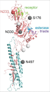
Structure of the ectodomain of a HEF monomer from IDV. The polypeptide chains of HEF1 and HEF2 subunits are colored in red and blue, respectively. The amino acids of the receptor binding site are marked as green sticks, the catalytic triad of the esterase domain as blue sticks. The location of asparagine residues of N-glycosylation sites (Asn-X-Ser/Thr) that are conserved between ICV and IDV HEF are marked as light gray balls. (One further conserved but unused site is not present in the crystalized ectodomain of HEF). Glycosylation sites not conserved between ICV and IDV HEF are marked as dark gray balls. S176 is a used glycosylation site in ICV HEF which is not present in IDV HEF. N330 and N497 are 2 glycosylation sites present in IDV, but not ICV HEF. The consensus sequence of the 27 HEF sequences in the database revealed another glycosylation site NKT at N233 (marked as red ball), that is changed to NKA in the HEF used for crystallization. Numbering starts with the first amino acid present in the mature protein, assuming signal peptide cleavage after residues 16. The figure was created with PyMol from PDB file 5e64, rendering was done with Blender.
Figure 5.
Figure 5.
Comparison of the receptor-binding sites of ICV and IDV HEF. Surface presentations of the receptor-binding site of HEF from ICV (A) and IDV (B). The parts of the HEF1 molecule involved in receptor-binding (the 170 loop, the 230 helix, the 270 and 290 loops) are colored yellow. The 5 residues F127 (C HEF: Y127), W185 (C HEF: L184), Y231 (C HEF: Y227), F229 (C HEF: F225) and F297 (C HEF: F293) that form the bottom of the cavity are colored red. Note that K235 (blue) and D269 (red) form a salt bridge in ICV HEF that connects helix 230 with loop 270. At equivalent positions of IDV HEF A273 and T239 (colored red) are present that are not able to form a salt bridge. As a result, the receptor cavity of IDV HEF is more open and thus might allow binding of more 9-O-Ac-Neu5Ac derivatives. The figure was created with PyMol from PDB files 5e64 and1flc and rendering was done with Blender.

Similar articles

Cited by

References

    1. Sandbulte MR, Westgeest KB, Gao J, Xu X, Klimov AI, Russell CA, Burke DF, Smith DJ, Fouchier RA, Eichelberger MC. Discordant antigenic drift of neuraminidase and hemagglutinin in H1N1 and H3N2 influenza viruses. Proceedings of the National Academy of Sciences of the United States of America. 2011;108:20748-53. doi:10.1073/pnas.1113801108. PMID:22143798 - DOI - PMC - PubMed
    1. Yoon SW, Webby RJ, Webster RG. Evolution and ecology of influenza A viruses. Current topics in microbiology and Immunology. 2014;385:359-75. PMID:24990620 - PubMed
    1. Olsen B, Munster VJ, Wallensten A, Waldenstrom J, Osterhaus AD, Fouchier RA. Global patterns of influenza a virus in wild birds. Science. 2006;312:384-8. doi:10.1126/science.1122438. PMID:16627734 - DOI - PubMed
    1. Watanabe T, Watanabe S, Maher EA, Neumann G, Kawaoka Y. Pandemic potential of avian influenza A (H7N9) viruses. Trends in Microbiology. 2014;22:623-31. doi:10.1016/j.tim.2014.08.008. PMID:25264312 - DOI - PMC - PubMed
    1. Lakdawala SS, Jayaraman A, Halpin RA, Lamirande EW, Shih AR, Stockwell TB, Lin X, Simenauer A, Hanson CT, Vogel L, et al.. The soft palate is an important site of adaptation for transmissible influenza viruses. Nature. 2015;526:122-5. doi:10.1038/nature15379. PMID:26416728 - DOI - PMC - PubMed

Publication types

MeSH terms