iBet uBet web content aggregator. Adding the entire web to your favor.


Link to original content: https://www.ncbi.nlm.nih.gov/pubmed/20627072
Tumors as organs: complex tissues that interface with the entire organism - PubMed Skip to main page content
U.S. flag

An official website of the United States government

Dot gov

The .gov means it’s official.
Federal government websites often end in .gov or .mil. Before sharing sensitive information, make sure you’re on a federal government site.

Https

The site is secure.
The https:// ensures that you are connecting to the official website and that any information you provide is encrypted and transmitted securely.

Access keys NCBI Homepage MyNCBI Homepage Main Content Main Navigation
Review
. 2010 Jun 15;18(6):884-901.
doi: 10.1016/j.devcel.2010.05.012.

Tumors as organs: complex tissues that interface with the entire organism

Affiliations
Review

Tumors as organs: complex tissues that interface with the entire organism

Mikala Egeblad et al. Dev Cell. .

Abstract

Solid tumors are not simply clones of cancer cells. Instead, they are abnormal organs composed of multiple cell types and extracellular matrix. Some aspects of tumor development resemble processes seen in developing organs, whereas others are more akin to tissue remodeling. Some microenvironments, particularly those associated with tissue injury, are favorable for progression of mutant cells, whereas others restrict it. Cancer cells can also instruct surrounding tissues to undergo changes that promote malignancy. Understanding the complex ways in which cancer cells interact with their surroundings, both locally in the tumor organ and systemically in the body as a whole, has implications for effective cancer prevention and therapy.


Figures

Figure 1
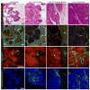
Figure 1. Changes in stromal organization during tumor progression
(A) Changes in overall tissue composition with tumor stages in the MMTV-PyMT mouse mammary carcinoma model. Hematoxylin and eosin stained sections (H&E). With tumor progression, the architecture of the carcinoma cells bears less and less resemblance to the architecture of the tissue from which it was derived. The stromal tissue also changes, for example, from a tissue dominated by adipocytes to one dominated by extracellular matrix, fibroblasts and immune cells (adapted from Egeblad et al., 2008; Lin et al., 2003). (B) Changes in extracellular matrix. Fibrillar collagens are stained with picrosirius red, shown as the birefringent stain under polarized light (arrows); counterstained with hematoxylin. With tumor progression, fibrillar collagen accumulates at the invasive edge of the tumor and surrounding nests of cancer cells (adapted from Levental et al., 2009; Provenzano et al., 2006). (D) With increasing tumor stage, the tumor vasculature becomes increasingly abnormal, e.g., with dilated vessels. The vasculature is labeled by intravenous injection of FITC-tomato lectin, green (arrows). Nuclei stained with propidium iodide are shown in red. Scale bars: 50 μm (adapted from Lin et al., 2006). (D) Leukocyte infiltration increases with tumor progression. Immune cells are labeled with antibodies against the common leukocyte marker CD45, green (arrows). Nuclei stained with propidium iodide are shown in blue (adapted from Egeblad et al., 2008).
Figure 2
Figure 2. Interactions between cancer cells and stromal components influence tumor growth and progression
(A) Interactions with mesenchymal cells. In hyperplastic tissues, normal epithelium and fibroblasts may exert a tumor-inhibiting effect. As cancer cells begin to expand, they also produce factors that activate myofibroblasts and recruit carcinoma-associated fibroblasts. These mesenchymal cell types, as well as adipocytes, are responsible for many of the tumor-associated changes in extracellular matrix (ECM). (B) Recruitment of cells of the innate and adaptive immune compartment to a carcinoma. The immune cells are found both in the stroma at the invading edge of the carcinoma and infiltrating the tumor. Inflammatory cells, including neutrophils and macrophages, are frequently the first immune cells recruited to the tumor, and may be either tumor-promoting or tumor-inhibiting depending on their polarization. Another inflammatory cell type, the mast cell, is also recruited early and promotes tumor progression by releasing proteases that activate angiogenesis. Dendritic cells are primarily tumor-inhibiting as they support immunosurveillance and release signals that activate cytotoxic T cells. In contrast, myeloid-derived suppressor cells function to inhibit T cell activation. Natural killer cells and different types of T cells may have either pro- or anti-tumor functions, depending on their mode of activation. Immunoglobulins released by B cells promote tumor growth by initiation of the inflammatory response. (C) Formation of metastases. At sites of vascular leakage, fibronectin is deposited and vascular endothelial growth factor receptor 1 (VEGFR1)-positive bone marrow-derived cells exit the circulation where they promote the establishment of the future metastases. They are involved in angiogenesis at the metastatic site through secretion of VEGF and degradation of the ECM by the release of MMP9. Circulating cancer cells reach the pre-metastatic site, sometimes covered by activated platelets, which protect them while they are in blood vessels and facilitate adhesion to the endothelial wall at the secondary site.
Figure 3
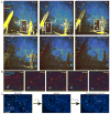
Figure 3. Dynamics of the interactions between cancer cells and stromal components in the tumor tissue
(A) Dynamics of different subtypes of myeloid cells revealed by four-color time-lapse series of the same lesion of an MMTV-PyMT;ACTB-ECFP;c-fms-EGFP mouse co-injected with fluorescent 70 kD dextran and anti-Gr1 antibodies intravenously. Dextran (yellow) first labels the blood vessels (0–6 h post injection), but leaks out of the vessels over time and is taken up by M2-like macrophages (3–12 h). The anti-Gr1 antibody labels Gr1+ myeloid cells, including neutrophils, myeloid derived suppressor cells and monocytes (red, arrow heads). Inserts show higher magnifications of blood vessels without the dextran channel for identification of Gr1+ myeloid cells at early time points. All myeloid cells are green and cancer cells are blue (adapted from Egeblad et al., 2008). See also Movie S1 in supplementary material. Scale bars: 100 μm. (B) Examples of low-migratory (white outline) and high-migratory (pink outline) CXCR6+ T-cells (green) in proximity to the tumor vasculature (red) in a live MMTV-PyMT;ACTB-ECFP;CXCR6EGFP mouse injected with fluorescent dextran (red). Cancer cells are blue (adapted from Egeblad et al., 2008). See also Movie S2 in supplementary material for time-lapse recordings of the entire field. (C) Effects of acute hypoxia on T cell migration. CXCR6+ T cells (green) cease migration following acute systemic hypoxia in a live MMTV-PyMT;ACTB-ECFP;CXCR6EGFP mouse. Cancer cells are blue. The same 15 cells were tracked during 20 minutes of normoxia (21% inhaled oxygen), 20 minutes of systemic acute hypoxia (7% inhaled oxygen) and 20 minutes of re-established normoxia in the same field (tracking marks are white; adapted from Egeblad et al., 2008). See also Movie S3 in supplementary material.
Figure 4
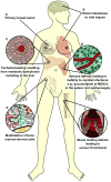
Figure 4. Systemic changes resulting from cancer
The interactions between the tumor and the rest of the body are primary causes of cancer-related morbidity and mortality. (A, B) Malignant cells from the primary tumor disseminate into circulation and colonize and expand into vital organs such as the lungs and brain. (C) Tumor-secreted factors, such as TNF-α and IFN-γ, can disrupt the normal metabolic functions of the liver and lead to cachexia, a wasting away of body mass. (D) The spleen has a major function in the mounting of immune responses. During cancer progression, myeloid-derived suppressor cells (MDSCs) accumulate, contributing to splenomegaly and an impaired immune response with increased susceptibility to infection. (E) In the bone marrow, mobilization of bone marrow-derived cells is increased. These cells promote tumor progression by activating angiogenesis and increasing invasiveness of cancer cells. (F) The primary tumors often activate pro-coagulatory factors and inhibit fibrinolytic factors, resulting in thrombosis formation. Clotting of the vasculature can be a sign of undiagnosed cancer, and is a major complication of late-stage cancer.
Figure 5
Figure 5. Tumor tissue organization influences drug response
(A) The overall architecture of the tumor has a direct effect on the ability of a cancer drug to penetrate the tissue and reach the cancer cells. Abnormal leakage from blood vessels together with insufficient lymphatic drainage, especially from the middle of tumors, contribute to increased interstitial pressure in the tumor tissue that inhibits penetration of drugs into the deeper areas of the tumors. Cancer drug penetration is also limited by their binding to ECM proteins, such as collagen, or their uptake by stromal cells, such as macrophages. Cancer cells furthest away from the blood vessel not only are exposed to the lowest drug concentrations, but also receive the lowest amounts of nutrients and oxygen from the circulation and therefore have the lowest proliferative index. As many cancer cell drugs preferentially target actively proliferating cells, this effect contributes to the inability of drugs to target cells in hypoxic areas. Cancer cells further from the blood vessel are also exposed to a low pH microenvironment where many cytotoxic drugs become inactive. Thus, the organization of the tumor tissue results in limited drug availability and efficacy in the hypoxic areas of tumors, areas speculated to contain some of the most aggressive cancer cells. The changing microenvironment in different parts of the tumors therefore results in an apparent difference in drug sensitivity (indicated by the changing color of the cancer cells). (B) Distribution of doxorubicin in vivo. Section from a mouse mammary tumor showing the distribution of doxorubicin (blue) in relation to tumor blood vessels (red) and regions of hypoxia (green). Note that doxorubicin is found only in close proximity to tumor blood vessels. Scale bar: 100 μm. Reprinted with permission from Primeau et al., 2005.

Similar articles

Cited by

References

    1. Acharyya S, Ladner KJ, Nelsen LL, Damrauer J, Reiser PJ, Swoap S, Guttridge DC. Cancer cachexia is regulated by selective targeting of skeletal muscle gene products. J Clin Invest. 2004;114:370–378. - PMC - PubMed
    1. Anderson AR, Weaver AM, Cummings PT, Quaranta V. Tumor morphology and phenotypic evolution driven by selective pressure from the microenvironment. Cell. 2006;127:905–915. - PubMed
    1. Andreu P, Johansson M, Affara NI, Pucci F, Tan T, Junankar S, Korets L, Lam J, Tawfik D, Denardo DG, et al. FcRgamma activation regulates inflammation-associated squamous carcinogenesis. Cancer Cell. 2010;17:121–134. - PMC - PubMed
    1. Apetoh L, Ghiringhelli F, Tesniere A, Obeid M, Ortiz C, Criollo A, Mignot G, Maiuri MC, Ullrich E, Saulnier P, et al. Toll-like receptor 4-dependent contribution of the immune system to anticancer chemotherapy and radiotherapy. Nat Med. 2007;13:1050–1059. - PubMed
    1. Ausprunk DH, Folkman J. Migration and proliferation of endothelial cells in preformed and newly formed blood vessels during tumor angiogenesis. Microvasc Res. 1977;14:53–65. - PubMed

Publication types

MeSH terms

LinkOut - more resources