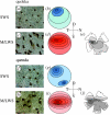
Cone topography and spectral sensitivity in two potentially trichromatic marsupials, the quokka (Setonix brachyurus) and quenda (Isoodon obesulus)
- PMID: 15888411
- PMCID: PMC1599861
- DOI: 10.1098/rspb.2004.3009
Cone topography and spectral sensitivity in two potentially trichromatic marsupials, the quokka (Setonix brachyurus) and quenda (Isoodon obesulus)
Abstract
The potential for trichromacy in mammals, thought to be unique to primates, was recently discovered in two Australian marsupials. Whether the presence of three cone types, sensitive to short- (SWS), medium- (MWS) and long- (LWS) wavelengths, occurs across all marsupials remains unknown. Here, we have investigated the presence, distribution and spectral sensitivity of cone types in two further species, the quokka (Setonix brachyurus) and quenda (Isoodon obesulus). Immunohistochemistry revealed that SWS cones in the quokka are concentrated in dorso-temporal retina, while in the quenda, two peaks were identified in naso-ventral and dorso-temporal retina. In both species, MWS/LWS cone spatial distributions matched those of retinal ganglion cells. Microspectrophotometry (MSP) confirmed that MWS and LWS cones are spectrally distinct, with mean wavelengths of maximum absorbance at 502 and 538 nm in the quokka, and at 509 and 551 nm, in the quenda. Although small SWS cone outer segments precluded MSP measurements, molecular analysis identified substitutions at key sites, accounting for a spectral shift from ultraviolet in the quenda to violet in the quokka. The presence of three cone types, along with previous findings in the fat-tailed dunnart and honey possum, suggests that three spectrally distinct cone types are a feature spanning the marsupials.
Figures


Similar articles
-
Topographies of retinal cone photoreceptors in two Australian marsupials.Vis Neurosci. 2003 May-Jun;20(3):307-11. doi: 10.1017/s0952523803203096. Vis Neurosci. 2003. PMID: 14570252
-
Trichromacy in Australian marsupials.Curr Biol. 2002 Apr 16;12(8):657-60. doi: 10.1016/s0960-9822(02)00772-8. Curr Biol. 2002. PMID: 11967153
-
Diversity of color vision: not all Australian marsupials are trichromatic.PLoS One. 2010 Dec 6;5(12):e14231. doi: 10.1371/journal.pone.0014231. PLoS One. 2010. PMID: 21151905 Free PMC article.
-
S cones: Evolution, retinal distribution, development, and spectral sensitivity.Vis Neurosci. 2014 Mar;31(2):115-38. doi: 10.1017/S0952523813000242. Epub 2013 Jul 29. Vis Neurosci. 2014. PMID: 23895771 Review.
-
[Current views on vision of mammals].Zh Obshch Biol. 2012 Nov-Dec;73(6):418-34. Zh Obshch Biol. 2012. PMID: 23330397 Review. Russian.
Cited by
-
The evolutionary history and spectral tuning of vertebrate visual opsins.Dev Biol. 2023 Jan;493:40-66. doi: 10.1016/j.ydbio.2022.10.014. Epub 2022 Nov 9. Dev Biol. 2023. PMID: 36370769 Free PMC article. Review.
-
Evolutionary Constraint on Visual and Nonvisual Mammalian Opsins.J Biol Rhythms. 2021 Apr;36(2):109-126. doi: 10.1177/0748730421999870. Epub 2021 Mar 25. J Biol Rhythms. 2021. PMID: 33765865 Free PMC article.
-
Evolution, Development and Function of Vertebrate Cone Oil Droplets.Front Neural Circuits. 2017 Dec 8;11:97. doi: 10.3389/fncir.2017.00097. eCollection 2017. Front Neural Circuits. 2017. PMID: 29276475 Free PMC article. Review.
-
The evolution of eyes: major steps. The Keeler lecture 2017: centenary of Keeler Ltd.Eye (Lond). 2018 Feb;32(2):302-313. doi: 10.1038/eye.2017.226. Epub 2017 Oct 20. Eye (Lond). 2018. PMID: 29052606 Free PMC article.
-
Euarchontan Opsin Variation Brings New Focus to Primate Origins.Mol Biol Evol. 2016 Apr;33(4):1029-41. doi: 10.1093/molbev/msv346. Epub 2016 Jan 6. Mol Biol Evol. 2016. PMID: 26739880 Free PMC article.
References
-
- Ahnelt K.P, Kolb H. The mammalian photoreceptor mosaic-adaptive design. Prog. Retin. Eye Res. 2000;19:711–777. - PubMed
-
- Arrese C.A, Hart N.S, Thomas N, Beazley L.D, Shand J. Trichromacy in Australian marsupials. Curr. Biol. 2002;12:657–660. - PubMed
-
- Arrese C.A, Rodger J, Beazley L.D, Shand J. Topographies of retinal cone photoreceptors in two Australian marsupials. Vis. Neurosci. 2003;20:303–311. - PubMed
-
- Beazley L.D, Dunlop S.A. The evolution of the area centralis and visual streak in the marsupial Setonix brachyurus. J. Comp. Neurol. 1983;216:211–231. - PubMed
-
- Bowmaker J.K. Evolution of photoreceptors and visual pigments. In: Cronly-Dillon J.R, Gregory R.L, editors. In Evolution of the eye and visual pigments. CRC Press; Boca Raton, FL: 1991. pp. 63–81.
Publication types
MeSH terms
Substances
LinkOut - more resources
Full Text Sources
Research Materials


