Abstract
Myiasis is derived from the Greek word-“Myia”, meaning “fly”. The term was first introduced by Hope in 1840 and refers to the infestation of human beings with dipterous larvae (maggots). Presence of maggots on exposed parts is already known, but on covered parts like external genitalia it is very rare. We hereby describe a case of young unmarried female who presented with multiple sinuses over external genitalia along with maggots coming out of it.
Keywords: External genitalia, female, maggots, myiasis, sinuses
INTRODUCTION
Myiasis is derived from the Greek word-"Myia", meaning “fly”. The term was first introduced by Hope in 1840 and refers to the infestation of human beings with dipterous larvae (maggots). Myiasis is the infestation of dipterous larva in humans and other vertebrate animals.[1] The larva feed on the host's dead or living tissues, liquid body substances or ingested food. Single female fly deposits 40-80 first stage larva, which are capable of penetrating tissue and thus cause immediate problems depending on the body site involved.[2] Maggots are seen on the exposed body parts with infected skin lesions in small neglected children, very old patients, mentally retarded, bedridden patients who are not able to take care of themselves. In young, healthy mentally sound and active persons it is rare to see maggots and too rare to be seen in covered parts like genitals.
CASE REPORT
A 17-year-old young unmarried female presented with a history of pain and swelling in the genitalia and also complaining of dropping of fly larva from vulva. She lived in the rural area with conditions of poor hygiene, which were compatible with high-risk of disease. Her non-hygienic toilet was outside the house and attracted many flies. Her hygiene was poor and wearing dirty clothes. She was accompanied by her aunt as her mother died in childhood and because of her shy nature, she didn’t report to any one until and unless she started having pain. There was a history of normal regular menstrual cycle prior to the onset of this painful swelling of genitalia. Patient did not use readymade sanitary napkins available in the market during the menstrual periods; instead, she used dirty ragged clothes during menstrual cycles. She used to hang the washed clothes and undergarments on a clothes line outside. There was no history of any trauma, insect bite, and sexual activity. There was no history of pain in the lower abdomen. No history of intake of any immunosuppressive or steroidal therapy. She also denied history of any chronic illness including tuberculosis, chronic urinary tract infection, and diabetes. Physical examination of the patient revealed a normal build (height 5 feet 2 inches, weight 52 kg) with vitals in normal range. General physical examination revealed that the patient was of sound physical and mental health and capable of caring herself. She was studying in 11th standard.
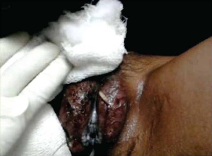
On local examination, both labia majora were tender, erythematous and swollen with the multiple discharging sinuses stuffed with crawling maggots of creamy-white color [Figure 1]. Labia minora were normal. Hymen was intact. No significant lymphadenopathy was present. Laboratory investigations revealed hemoglobin-9.8 g%, with a normal total and differential leucocytes count, blood sugar-98 g%, urine complete examination-within normal limits, her serology for HIV (Human immunodeficiency virus) and Syphilis (by Venereal Disease Research Laboratory test) was -Negative. Her urine examination and sonography was negative for pregnancy. She was hospitalized and was given injection ceftriaxone, injection metrogyl, injection gentamycin, tablet serratiopeptidase and tablet cetrizine empirically from the 1st day. Initially, on day 1, about 20 maggots were removed using non-toothed forceps and the wound was cleaned with betadine. On 2nd day, we applied Turpentine oil and more maggots were removed on 2nd and 3rd day. On the 4th day, maggots were completely absent. Lesions healed within a week time, patient was discharged and advised regarding personal hygiene to avoid re-infestation.
Figure 1.

Multiple discharging sinuses over the labia majora and maggot coming out of left labia majora sinus
DISCUSSION
The word maggots means larva of the fly. It is the non-technical word and the technical term is myiasis, which is defined as a disease caused by the infiltration of body tissue by house fly's larva.[3] Maggots like fly larva are of wide importance in ecology, economy, surgery, and forensic medicine. Myiasis occurs predominantly in rural areas with poor hygiene and low education level.[4] Maggots can enter through intact skin or through a wound. They may also enter a body orifice without tissue invasion (pseudo-myiasis).
The classical description of myiasis is according to the part of the host that is infected.[5]
Dermal
Sub-dermal
- Cutaneous
- Creeping, where larva burrow through or under the skin
- Furuncular, where a larva remains in one spot, causing a boil-like lesion
Nasopharyngeal nose, sinuses or pharynx
Ophthalmic or ocular in or about the eye
Auricular in or about the ear
Gastric, rectal or intestinal/enteric for the appropriate part of the digestive system
Urogenital.
Vulvar myiasis is a rare entity and constitutes only 0.7% of human infestation.[6] We consider our patient as a case of myiasis as the maggots have invaded the vulvar tissue (labia majora). As poor hygiene is known to be associated with vulvar myiasis, washing, and keeping the genital area clean may prevent the occurrence of this condition to a great extent.[7,8] The possible source in the present case may be the eggs, which were transmitted to the vulva via the soiled clothes. It is a routine habbit among villagers to dry their washed cloths on ground. The flies laid eggs when the undergarment was on clothes line as flies were attracted to the blood and body secretions. In a multi-center study of American patients who were infected with maggots, 42 cases of myiasis were identified with male predominance and 38% were homeless.[6] Sherman recommended that maggots should be sent for species identification.[7] Unfortunately, species identification was not possible because of lack of the facility in the nearby area. Many authors believed that vulvar myiasis remains under reported and they recommended that it should be reported accordingly.[4] The use of turpentine oil has been advocated in many cases of cutaneous myiasis world-wide, but none have mentioned its use in vulvar myiasis.[9] The use of turpentine oil produced excellent results in our case. In our case, no pre-existing genital lesion and sero-positivity to HIV was present as a precipitating cause of myiasis.[7,10,11,12] The lack of personal hygiene is the contributing factor for the cause of myiasis, more so with the genital myiasis. Baidya has reported a case of genital myiasis in women with genital prolapse and malignancy.[13] Passos et al. reported a case of vulvar myiasis during the pregnancy.[4] Cilla et al. described a case of vulvar myiasis in a diabetic 86 year old women.[14]
To conclude, myiasis in external genitalia is a rare condition. This case highlights the need for thorough genital examination as a means of identifying less common disease. The physician's role in educating patients (especially those living in a rural area) is important and must emphasize the role of good personal hygiene.
Footnotes
Source of Support: Nil
Conflict of Interest: None declared.
REFERENCES
- 1.Robbins K, Khachemoune A. Cutaneous myiasis: A review of the common types of myiasis. Int J Dermatol. 2010;49:1092–8. doi: 10.1111/j.1365-4632.2010.04577.x. [DOI] [PubMed] [Google Scholar]
- 2.Burgess IF. Myiasis: Maggots infestation. Nurse Times. 2003;99:51–3. [PubMed] [Google Scholar]
- 3.Mandell GL, Bennett JE, Dolin R. Myiasis. In: Mandell GL, Douglas RG, Bennett JE, editors. Principles and Practice of Infectious Diseases. Philadelphia: Churchill Livingstone; 2000. p. 2976. [Google Scholar]
- 4.Passos MR, Varella RQ, Tavares RR, Barreto NA, Santos CC, Pinheiro VM, et al. Vulvar myiasis during pregnancy. Infect Dis Obstet Gynecol. 2002;10:153–8. doi: 10.1155/S1064744902000157. [DOI] [PMC free article] [PubMed] [Google Scholar]
- 5.Janovy J, Schmidt & Larry S, Gerald D. Schmidt & Larry S. Roberts’ Foundations of Parasitology. In: Roberts S, Larry S, Gerald D, editors. Dubuque, Iowa: Wm. C. Brown; 1996. ISBN 0-697-26071-2. [Google Scholar]
- 6.Sherman RA. Wound myiasis in urban and suburban United States. Arch Intern Med. 2000;160:2004–14. doi: 10.1001/archinte.160.13.2004. [DOI] [PubMed] [Google Scholar]
- 7.Predy G, Angus M, Honish L, Burnett CE, Stagg A. Myiasis in an urban setting: A case report. Can J Infect Dis. 2004;15:51–2. doi: 10.1155/2004/978427. [DOI] [PMC free article] [PubMed] [Google Scholar]
- 8.Yazar S, Ozcan H, Dinçer S, Sahin I. Vulvar myiasis. Yonsei Med J. 2002;43:553–5. doi: 10.3349/ymj.2002.43.4.553. [DOI] [PubMed] [Google Scholar]
- 9.Sharma J, Mamatha GP, Acharya R. Primary oral myiasis: A case report. Med Oral Patol Oral Cir Bucal. 2008;13:E714–6. [PubMed] [Google Scholar]
- 10.Radotra A, Goel P, Thami GP, Aggrawal A, Wanchu M, Bhalla M. Vulvar myiasis complicating condylomata acuminate in pregnancy. Ind J Dermatol. 2004;49:207. [Google Scholar]
- 11.Dhawan J, Singh S, Gupta S. Insects are Crawling in My Genital Warts. J Cutan Aesthet Surg. 2011;4:129–31. doi: 10.4103/0974-2077.85037. [DOI] [PMC free article] [PubMed] [Google Scholar]
- 12.Ghosh SK, Bandyopadhyay D, Sarka S. Myiasis in a large perigenital seborrheic keratosis. Ind J Dermatol. 2010;55:305–6. doi: 10.4103/0019-5154.70699. [DOI] [PMC free article] [PubMed] [Google Scholar]
- 13.Baidya J. A rare case of genital myiasis in a woman with genital prolapse and malignancy and review of the literature. Ann Trop Med Public Health. 2009;2:29–30. [Google Scholar]
- 14.Cilla G, Picó F, Peris A, Idígoras P, Urbieta M, Pérez Trallero E. Human genital myiasis due to Sarcophaga. Rev Clin Esp. 1992;190:189–90. [PubMed] [Google Scholar]


