Abstract
Magnetic separation technology, using magnetic particles, is quick and easy method for sensitive and reliable capture of specific proteins, genetic material and other biomolecules. The technique offers an advantage in terms of subjecting the analyte to very little mechanical stress compared to other methods. Secondly, these methods are non-laborious, cheap and often highly scalable. Moreover, techniques employing magnetism are more amenable to automation and miniaturization. Now that the human genome is sequenced and about 30,000 genes are annotated, the next step is to identify the function of these individual genes, carrying out genotyping studies for allelic variation and SNP analysis, ultimately leading to identification of novel drug targets. In this post-genomic era, technologies based on magnetic separation are becoming an integral part of todays biology laboratory. This article briefly reviews the selected applications of magnetic separation techniques in the field of biotechnology, biomedicine and drug discovery.
Introduction
Magnetic fluids or ferrofluids as they are often called mainly consist of nano sized iron oxide particles (Fe3O4 or γ-Fe2O3) which are suspended in carrier liquid. In recent years, substantial progress has been made in developing technologies in the field of magnetic microspheres, magnetic nanospheres and ferrofluids. Techniques based on using magnetisable solid-phase supports (MSPS) have found application in numerous biological fields viz. diagnostics, drug targeting, molecular biology, cell isolation and purification, radio immuno assay, hyperthermia causing agents for cancer therapy, nucleic acid purification etc [1-3]. Although often referred to as magnetic, many of the particles currently used are superparamagnetic, meaning that these particles can be easily magnetized with an external magnetic field and redispersed immediately once the magnet is removed. Currently available formats of particles can be broadly classified into unmodified or naked particles, chemically derivatized particles with general specificity ligands (streptavidin, Protein A etc) and chemically derivatized particles with specific recognition groups viz. monoclonal and polyclonal antibodies [4]. This article discusses the selected advancement made in the field of biotechnology, medicine and drug discovery using magnetically driven separation techniques.
Drug discovery and genomics applications
The modern drug discovery process emphasizes rapid data generation and analysis to identify promising new chemical entities as well as new drug targets early in the development cycle. At every step of the rapidly evolving drug discovery process, dozens of technologies and products are required. But innovations in newer technologies for genomics and proteomics are changing the face of drug discovery. Automation has become essential in allowing researchers to meet the high through demands of today's research environment. The main thrust area where magnetic separation is applied in drug discovery is sample preparation that includes high throughput genome isolation for sequencing or PCR amplification to carry out genotyping, SNP scoring or expression profiling. The inherent benefits offered by magnetic handling includes, reduced reagent costs, elimination of labour intensive steps, easy automation and yield of high purity DNA in less amount of time compared to conventional methods.
Highthroughput DNA isolation
Isolation of DNA is a prerequisite step for many molecular biology techniques. The separation of DNA from the complex mixtures in which they are often found is frequently necessary before other studies and procedure like sequencing, amplification, hybridization, detection etc. The presence of large amounts of cellular or other contaminating material like proteins and RNA, in such complex mixtures often impedes many of the reactions and techniques used in molecular biology [5]. The conventional protocol for extracting DNA involves cell lysis followed by removal of contaminating cellular components such as proteins, lipids and carbohydrates; and finally isolating DNA using a series of precipitation and centrifugation steps, which are difficult to automate. Improvement in methods for isolating DNA has been made and more recently, methods that rely on the use of solid phase have been proposed. Adsorbents that provide fast, efficient DNA purification are important for making this procedure amenable to automation. The discovery in late 80's that silica can be used as adsorbent for DNA isolation [6], became the basis for most of the DNA isolation kits currently available. One of these kits involves isolation of DNA using silica coated magnetic particles [7].
A high throughput genome isolation protocol has been developed, which is based on SPRI (Solid Phase Reversible Immobilization) chemistry [8,9]. The SPRI protocol is based on DNA binding to the surface of carboxyl coated paramagnetic particles under the condition of high salt and PEG. The procedure has been developed and optimized for single stranded DNA isolation, such as M13 phage and double stranded plasmid DNA utilizing carboxyl coated magnetic particles. The SPRI protocol has allowed the development of an automated procedure in a microplate format with a throughput of about 200, 000 DNA preparations per day, thus becoming a fastest microplate plate based DNA purification system in use on the human genome sequencing.
Magnetic separation of poly (A) mRNA
A number of methods have been reported for isolation of total RNA from a variety of cells or tissues. Chirgwin et al. [10] developed a method for efficient isolation of total RNA by homogenization in a 4 M solution of guanidium thiocyanate containing 0.1 M 2-mercaptoethanol. Homogenization is followed by extraction of RNA by ethanol or by ultracentrifugation through cesium chloride. This method was further modified by Chomczynski and Sacchi [11] to devise a rapid single step isolation procedure for RNA. It involves extraction of RNA using a mixture of guanidium thiocyanate and phenol-chloroform. Many RNA isolation kits are available based on the above two protocols. All these methods isolate RNA on the basis of its biochemical properties. In contrast, biomagnetic separation of mRNA is based on specific complementary hybridization between poly A sequence of isolated mRNA and oligo (dT)25 sequence covalently linked to the surface of paramagnetic particles. In this method oligo (dT)25 coated magnetic beads are added to crude cell or tissue lysate. During incubation poly (A) mRNA from the lysate are caught onto the surface of oligo (dT)25 coated magnetic beads. The beads/mRNA complex is then washed magnetically. The mRNA thus isolated is either eluted or directly applied for many downstream applications which includes cDNA library construction, Substractive hybridization, Northern hybridization, RT-PCR and in vitro translation [12].
Isolation of nucleic acid binding biomolecules
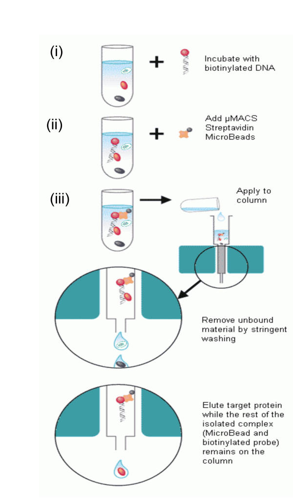
The isolation of specific molecules based on its interaction with complementary binding partner is emerging as important technologies in many field of research. The isolation and characterization of specific transcripts or proteins can be employed to monitor the progression of disease. Several kits are available in the market that works on the principle of magnetic labeling and direct isolation of biotinylated molecules such as DNA, RNA or proteins onto streptavidin coated magnetic beads (μMACS streptavidin Microbeads from Miltenyi Biotec, Germany). These biotinylated molecules can then be used for indirect isolation of non-biotinylated target molecules that may interact with them [13]. The procedure involves complex formation between the biotinylated probe (DNA, RNA or proteins) and the target molecule (i.e. interacting biomolecules DNA, RNA or protein). Based on the interaction of biotin-streptavidin, the probe-target complex is then separated from rest of the component by addition of streptavidin coated magnetic beads. The complex is magnetically isolated and washed to remove non-specifically bound molecules. The non-biotinylated target molecules can be either eluted off from the complex with high purity, whereas the magnetically labeled biotinylated probe remains bound to the column (Fig. 1). This technique has potential for rapid and efficient screening of transcriptional and translational regulatory proteins. Alam and co-workers have also used target DNA-conjugated magnetic beads for rapid screening of DNA-binding peptide ligands from solid phase combinatorial library [14].
Figure 1.
Isolation of nucleic acid binding molecules. Biotinylated DNA is added to the cell lysate or a protein mixture that contains protein which interacts with the DNA (i). Streptavidin magnetic beads are added that binds to the complex (biotin-DNA-protein) based on its affinity for biotin (ii). The complex is separated magnetically from the mixture (iii). Reprinted, with permission, from materials provided by Miltenyi Biotec, GmbH, Germany.
Proteomics Applications
Magnetocapture protein interaction assays
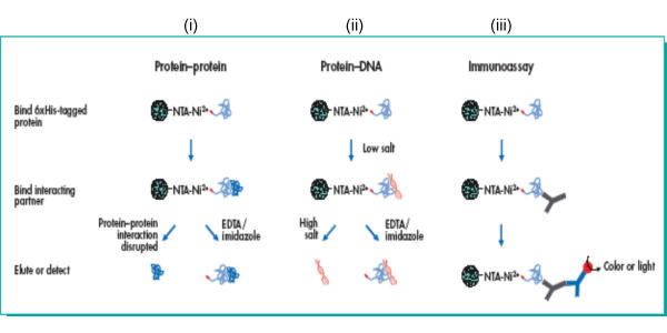
Magnetic particles are now increasingly used as carriers for binding proteins, enzymes and drugs. Studies have shown that proteins and enzymes can be bound covalently to naked magnetic particles in the presence of carbodiimide [15]. Such immobilization procedures for proteins, enzymes or drugs will have a major impact in various areas of medicine and biotechnology. The immobilized biomolecules can be used directly for a bioassay or as affinity ligands to capture or modify target molecules or cells. On this basis, Ni-NTA (nitriloacetic acid) tagged magnetic agarose beads have been used for versatile magnetocapture assays using 6xHis-tagged proteins [16]. The procedure involves use of metal chelating nitriloacetic acid (NTA) groups covalently bound to the surface of agarose beads, which contain strong magnetic particles. The beads are precharged with nickel, which is ready to capture 6xHis-tagged proteins for sensitive interaction assays or microscale purification of 6xHis tagged proteins (Fig. 2). Thus, this technique bridge the gap between purification-scale procedures using Ni-NTA metal chelate affinity chromatography resins and microplate-based assays [17]. Other procedures for magnetic affinity separations of various proteins have been summarized recently [18].
Figure 2.
Magnetocapture protein interaction assays. Ni-NTA magnetic agarose beads precharged with nickel captures 6xHis-tagged proteins, which can be used for studying protein-protein (i) and DNA-protein interactions (ii), and immunoassay procedures using antibodies specific for antigens present on the captured 6xHis-tagged biomolecule or antigens present on interaction partners bound to the captured 6xHis-tagged biomolecule (iii). Reprinted, with permission, from materials provided by Qiagen, GmbH, Germany.
Biomedical Applications
Drug Targeting Using Magnetic Targeted Carriers
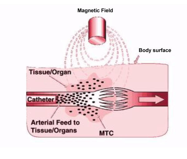
Magnetically guided drug targeting has been attempted in order to increase the efficacy and reduce the unpleasant side effects associated with chemotherapy. This method of drug delivery involves immobilization of drug or radionuclide in biocompatible magnetic nano- or microspheres [19]. This method of delivery makes chemotherapy more effective by increasing the drug concentration at the tumour site, while limiting the systemic drug concentration. FeRx Inc. of San Diego have designed Magnetic Targeted Carriers termed MTCs for site-specific targeting, tissue retention, and sustained release of drugs. These MTCs are composed of elemental iron particles and activated carbon. MTCs (1–2 μm in size) can adsorb and desorb pharmaceutical agents such as doxorubicin (DOX). Vialed MTCs are mixed with an anticancer drug already in solution; the mixture is then introduced into the catheter. The drug delivery using MTCs includes insertion of catheter into an arterial feed to the tumor, followed by application of powerful magnetic field to cause the MTC-DOX to extravasate through the capillary bed into the targeted tissue (Fig. 3). The field is left in place for another 15 minutes; after removal of the magnet, the particles remain trapped in the tumor, where the drug is then released [20]. FeRx has already initiated phase III clinical trial for MTC-DOX formulation [21]. Like wise, Hafeli and co-workers have carried out preclinical studies in collaboration with FeRx for treatment of liver and brain tumors using MTCs labeled with β-emitters such as Rhenium-188 and Yttrium-90 [22]. Although the main research concern of MTC-drug therapy is cancer treatment, the technology is not limited to this, other drugs that are deliverable in this manner include antibiotics, thrombolytics, anti-inflammatories, peptides and steroids.
Figure 3.
Drug targeting using MTCs. MTCs are composite of elemental iron and activated carbon with anticancer drug adsorbed onto it. MTCs are delivered intra-arterially via a catheter to the desired site by externally applied magnetic field. The magnetic field aids in particle localization and retention at targeted site by extravasation of the particles into the surrounding tissue. Reprinted, with permission, from materials provided by FeRx Inc., San Diego, CA.
Magnetoliposomes as drug delivery vehicles
Magnetoliposomes are magnetic derivatives of liposomes and can be prepared by entrapment of ferrofluids within the core of liposomes [23]. Several groups have investigated the uses of magnetoliposomes for site specific targeting [24], cell-sorting [25] and as magnetic resonance contrast enhancing agents [26].
Babincova et al. [27] have tried to use magnetoliposomes with encapsulated doxorubicin (DOX) for site specific chemotherapy in response to externally applied AC magnetic field. The result of this study showed that magnetoliposomes can be specifically heated to 42°C in a few minutes and during this, the encapsulated doxorubicin is massively released. Another in vivo studies for site specific targeting using magnetoliposomes incorporated with adriamycin (ADR), showed that administration of magnetic ADR liposomes under an applied external magnetic force produced approximately 4-fold higher maximum ADR concentration in the tumor than did administration of ADR solution alone. These results suggested that systemic chemotherapy could effectively control the primary tumor without significant side effects, due to the targeting of magnetic ADR liposomes [28,29].
Magnetotactic bacteria belonging mainly to Magnetospirillum species produces ferromagnetic crystalline particles consisting of magnetite (Fe3O4) or greigite (Fe3S4). These are called bacterial magnetic particles (BMPs). The BMPs are small in size (50–100 nm) and disperse very well because they are covered with a stable lipid membrane [30]. Magnetoliposomes containing cis-diamminedichloroplatinum (II) (CDDP) prepared using BMPs have also been evaluated for targeting and controlled release of drugs at tumor site. It has been observed from such studies that the capture volume of the magnetoliposomes with BMPs was 1.7 times higher than the ones with artificial magnetic particles. Also, when a rotating magnetic field was applied to these magnetoliposomes, the contents of them were released over 2 hours, suggesting that magnetoliposomes containing CDDP shows antitumor activity and that the controlled release of drugs from magnetoliposomes is possible [31].
Magnetic fluid hyperthermia
Magnetic fluids have been investigated as potential hyperthermia causing agents due to their high specific absorption rate (SAR). Hyperthermia is a promising approach for cancer treatment that uses AC magnetic fields to heat target areas (cancer tissue) containing magnetic fluids. To study the biological effects of AC magnetic field excited ferrofluids, both in-vitro and in-vivo studies have been carried out in cancer cell lines and spontaneously induced tumors in animal models. The results of these studies have shown that magnetic fluid hyperthermia is able to reduce the viability of cancer cells, thereby indicating the potential of this therapy [32]. There also exist the possibility of combination therapy, which would include hyperthermia treatment followed by chemotherapy or gene therapy. The approach involves use of magnetic nano- or microspheres or magnetoliposomes containing a drug to cause hyperthermia using the standard procedure, followed by the release of encapsulated drug that will act on the injured cells. It is anticipated that the combined treatment might be very efficient for treating solid tumor [19].
Cell Sorting Applications
Immunomagnetic cell isolation and separation
Immunomagnetic cell separation methods have become increasingly popular among the cell biologist. The general approach involves use of paramagnetic particles coated with antibodies against the target specific cell surface molecules. The method employs two schemes for isolating the target cell, direct method that involves coupling of affinity ligand (antibody) onto the magnetic particles, which are then directly added to the sample containing the target cells. During the incubation the magnetic particles binds the target cells that can then be recovered using a magnet. While in case of indirect method, a free affinity ligand (an appropriate antibody) is first added to the cell suspension. After incubation excess unbound affinity ligand is removed by washing, and the antibody-target cell complex is then captured by magnetic particles bearing an affinity ligand (secondary antibody) with the affinity for the primary label. Both positive and negative selection can be performed with immunomagnetic separation. In case of positive selection target cellular subsets are magnetically labeled and subsequently separated, while negative selection involves target purification by removing all other contaminating cells [33,34].
Using this technology large number of cell types has been isolated to date. Some of the examples where immunomagnetic separation technology has been applied includes, detection and removal of circulating tumor cells from peripheral blood [35], isolation of antigen specific CD 8+ T cells [36], selective separation of CD34+ cells [37], isolation and enrichment of retinal ganglion cells (RGCs) for culturing [38] etc. Immunomagnetic separation is used extensively for detection of pathogenic bacteria in clinical, food and environmental samples [33,39]. In addition to whole cell isolation, even cell organelles can be selectively separated using magnetic particles. The lysosome fraction was isolated from the amoeba Dictyostelium discoideum after feeding with dextran-based nanoparticles and subsequent homogenization [40]. Likewise a variety of cell types can be isolated using automated systems and different surface coated magnetic particles that are available from Miltenyi Biotec, Germany, or StemCell Technologies, USA.
Immunomagnetic assays
Immunoassays are becoming an important tool in clinical diagnostics and in fundamental investigations, because of its sensitivity, specificity and general applicability. Magnetic particles have been applied increasingly for various immunoassays that include fluoroimmunoassays, enzyme immunoassays or radioimmunoassays. Magnetic particles bound with primary or secondary antibodies are used for separation and quantification of antigens. Use of magnetically bound antibodies eliminates the centrifugation step thus reducing assay time and simplifying operation thereby increasing the efficiency and accuracy of the assay. Matsunaga and co-workers [41] have developed a novel fluoroimmunoassay method, where a fluorescein isothiocyanate (FITC) conjugated monoclonal anti-Escherichia coli antibody was immobilized onto bacterial magnetic particles (BMPs) for detection and removal of E. coli. The same group has also developed a chemiluminescence enzyme immunoassay with BMPs using IgG as a model antigen [42]. Likewise a magnetic bead based ELISA has been developed for detection of Staphylococcus species [43].
Conclusion
Magnetic separation technologies have been applied in diverse field of biology. This includes high throughput isolation of nucleic acids, proteins, cells or cell organelles. Now a days, several instruments are available from different companies that couples separation of biomolecules with its detection in terms of its quantification or its interactions with other biomolecules. This instruments either uses directly ferromagnetic particle as label (magneto assay) or couples magnetic particles with other detection methods such as fluorescence or chemiluminescence. It might be possible in near future that magnetic particles would be used as detection probes for a variety of assays, replacing labeling techniques such as fluorescence, chemiluminescence and radioactivity. In our lab, research work on similar lines is proposed. The work involves developing a detection method for bio-molecular interaction using magnetic particles as label. The method that is planned to be developed would have special emphasis in microarray technology, where bio-molecular interactions like cDNA-mRNA or DNA-DNA are the basis for determining gene expression or allelic variation. To conclude what can be said is, magnetism based separation technology holds immense potential in this post genomics era because of its high speed, low cost and amenability of automation.
Author's contributions
ZMS carried out the review of literature and drafted the manuscript, SDT and CNR did the necessary corrections to the manuscript. All authors read and approved the final manuscript.
Acknowledgments
Acknowledgement
The financial support to ZMS [F.NO. 9/114(131)/2K2/EMR-I] from Council of Scientific and Industrial Research (CSIR), New Delhi, India is gratefully acknowledged.
Contributor Information
ZM Saiyed, Email: zainul_s@yahoo.com.
SD Telang, Email: sdtelang@hotmail.com.
CN Ramchand, Email: cnramchand@yahoo.com.
References
- Ramchand CN, Priyadarshini P, Kopcansky P, Mehta RV. Application of magnetic fluids in medicine and biotechnology. Indian J Pure Appl Phys. 2001;39:683–686. [Google Scholar]
- Safarikova M, Safarik I. The application of magnetic techniques in biosciences. Magn Electr Sep. 2001;10:223–252. [Google Scholar]
- Safarik I, Safarikova M. Overview of magnetic separations used for biochemical and biotechnological applications. In: Hafeli U, Schutt W, Teller J, Zborowski M, editor. In Scientific and Clinical Applications of Magnetic Carriers. New York: Plenum Press; 1997. pp. 323–340. [Google Scholar]
- Sinclair B. To bead or not to bead: applications of magnetic bead technology. Scientist. 1998;12:17. [Google Scholar]
- Ahern H. Technology makes DNA isolation, purification simple and swift. Scientist. 1995;9:17. [Google Scholar]
- Vogelstein B, Gillespie D. Preparative and analytical purification of DNA from agarose. Proc Natl Acad Sci USA. 1979;76:615–619. doi: 10.1073/pnas.76.2.615. [DOI] [PMC free article] [PubMed] [Google Scholar]
- TECAN Inc http://www.tecan.com/la2000_dnaextraction_.pdf
- Hawkins TL, O'Connor-Morin T, Roy A, Santillan C. DNA purification and isolation using a solid-phase. Nucleic Acids Res. 1994;22:4543–4544. doi: 10.1093/nar/22.21.4543. [DOI] [PMC free article] [PubMed] [Google Scholar]
- Hawkins TL, McKernan KJ, Jacotot LB, MacKenzie JB, Richardson PM, Lander ES. A magnetic attraction to high-throughput genomics. Science. 1997;276:1887–1889. doi: 10.1126/science.276.5320.1887. [DOI] [PubMed] [Google Scholar]
- Chirgwin JM, Przybyla AE, MacDonald RJ, Rutter WJ. Isolation of biologically active ribonucleic acid from sources enriched in ribonuclease. Biochemistry. 1979;18:5294–9. doi: 10.1021/bi00591a005. [DOI] [PubMed] [Google Scholar]
- Chomczynski P, Sacchi N. Single step method of RNA isolation by guanidium thiocyanate-phenol chloroform extraction. Anal Biochem. 1987;162:156–9. doi: 10.1006/abio.1987.9999. [DOI] [PubMed] [Google Scholar]
- Mrazek F, Petrek M. Processing of mRNA from human leukocytes by biomagnetical separation: comparison with current methods of RNA isolation. Acta Univ Palacki Olomouc Fac Med. 1999;42:23–28. [PubMed] [Google Scholar]
- Albig A. Isolation of mRNA binding proteins using the μMACS streptavidin kit. MACS & more. 2001;5:6–7. [Google Scholar]
- Alam MR, Maeda M, Sasaki S. DNA-binding peptides searched from the solid-phase combinatorial library with the use of the magnetic beads attaching the target duplex DNA. Bioorg Med Chem. 2000;8:465–73. doi: 10.1016/s0968-0896(99)00298-9. [DOI] [PubMed] [Google Scholar]
- Koneracka M, Kopcansky P, Timko M, Ramchand CN, de Sequeira A, Trevan M. Direct binding procedure of proteins and enzymes to fine magnetic particles. J Mol Catal B – Enzym. 2002;689:1–6. [Google Scholar]
- Sinclair B. Honing your cloning: new cloning systems give protein expression studies a boost. Scientist. 2000;14:29. [Google Scholar]
- QIAGEN Inc http://www.qiagen.com/literature/qiagennews/0498/984ninta.pdf
- Safarik I, Safarikova M. Biologically active compounds and xenobiotics: magnetic affinity separations. In: Wilson ID, Adlard TR, Poole CF, Cool M, editor. In Encyclopedia of Separation Science. London: Academic Press; 2000. pp. 2163–2170. [Google Scholar]
- Safarik I, Safarikova M. Magnetic nanoparticles and biosciences. Mon Chem. 2002;133:737–759. [Google Scholar]
- Fricker J. Drugs with a magnetic attraction to tumours. Drug Discov Today. 2001;6:387–89. doi: 10.1016/s1359-6446(01)01771-8. [DOI] [PubMed] [Google Scholar]
- FeRx Inc http://www.ferx.com/004trial02june031.htm
- Schutt W, Gruttner C, Teller J, Westphal F, Hafeli U, Paulke B, Goetz P, Finck W. Biocompatible magnetic polymer carriers for in vivo radionuclide delivery. Artif Organs. 1999;23:98–103. doi: 10.1046/j.1525-1594.1999.06278.x. [DOI] [PubMed] [Google Scholar]
- De Cuyper, Joniau M. Magnetoliposomes: formation and structural characterization. Eur Biophys J. 1988;15:311–319. doi: 10.1007/BF00256482. [DOI] [PubMed] [Google Scholar]
- Babincova M, Altanerova V, Lampert M, Altaner C, Machova E, Sramka M, Babinec P. Site-specific in vivo targeting of magnetoliposomes using externally applied magnetic field. Z Naturforsch (C) 2000;55:278–281. doi: 10.1515/znc-2000-3-422. [DOI] [PubMed] [Google Scholar]
- Margolis LB, Namiot VA, Kljukin LM. Magnetoliposomes: another principle of cell sorting. Biochim Biophys Acta. 1983;735:193–195. doi: 10.1016/0005-2736(83)90276-6. [DOI] [PubMed] [Google Scholar]
- Bulte JW, Cuyper MD, Despres D, Frank JA. Short- vs. long-circulating magnetoliposomes as bone marrow-seeking MR contrast agents. J Magn Reson Imaging. 1999;9:329–335. doi: 10.1002/(sici)1522-2586(199902)9:2<329::aid-jmri27>3.0.co;2-z. [DOI] [PubMed] [Google Scholar]
- Babincova M, Cicmanec P, Altanerova V, Altaner C, Babinec P. AC-magnetic field controlled drug release from magnetoliposomes: design of a method for site-specific chemotherapy. Bioelectrochemistry. 2002;55:17–9. doi: 10.1016/s1567-5394(01)00171-2. [DOI] [PubMed] [Google Scholar]
- Kubo T, Sugita T, Shimose S, Nitta Y, Ikuta Y, Murakami T. Targeted delivery of anticancer drugs with intravenously administered magnetic liposomes in osteosarcoma-bearing hamsters. Int J Oncol. 2000;17:309–15. doi: 10.3892/ijo.17.2.309. [DOI] [PubMed] [Google Scholar]
- Kubo T, Sugita T, Shimose S, Nitta Y, Ikuta Y, Murakami T. Targeted systemic chemotherapy using magnetic liposomes with incorporated adriamycin for osteosarcoma in hamsters. Int J Oncol. 2001;18:121–125. doi: 10.3892/ijo.18.1.121. [DOI] [PubMed] [Google Scholar]
- Schuler D, Frankel RB. Bacterial magnetosomes: microbiology, biomineralization and biotechnology applications. Appl Microbiol Biotech. 1999;52:464–473. doi: 10.1007/s002530051547. [DOI] [PubMed] [Google Scholar]
- Matsunaga T, Higashi Y, Tsujimura N. Drug delivery by magnetoliposomes containing bacterial magnetic particles. Cell Eng. 1997;2:7–11. [Google Scholar]
- Jordan A, Scholz R, Wust P, Fahling H, Krause J, Wlodarczyk W, Sander B, Vogl T, Felix R. Effects of magnetic fluid hyperthermia (MFH) on C3H mammary carcinoma in vivo. Int J Hyperthermia. 1997;13:587–605. doi: 10.3109/02656739709023559. [DOI] [PubMed] [Google Scholar]
- Safarik I, Safarikova M. Use of magnetic techniques for the isolation of cells. J Chromatogr B Biomed Sci Appl. 1999;722:33–53. [PubMed] [Google Scholar]
- Safarik I, Safarikova M. Cells isolation: magnetic techniques. In: Wilson ID, Adlard TR, Poole CF, Cool M, editor. In Encyclopedia of Separation Science. London: Academic Press; 2000. pp. 2260–2267. [Google Scholar]
- Bilkenroth U, Taubert H, Riemann D, Rebmann U, Heynemann H, Meye A. Detection and enrichment of disseminated renal carcinoma cells from peripheral blood by immunomagnetic cell separation. Int J Cancer. 2001;92:577–82. doi: 10.1002/ijc.1217. [DOI] [PubMed] [Google Scholar]
- Luxembourg AT, Borrow P, Teyton L, Brunmark AB, Peterson PA, Jackson MR. Biomagnetic isolation of antigen-specific CD8+ T cells usable in immunotherapy. Nat Biotechnol. 1998;16:281–285. doi: 10.1038/nbt0398-281. [DOI] [PubMed] [Google Scholar]
- Kato K, Radbruch A. Isolation and characterization of CD34+ hematopoietic stem cells from human peripheral blood by high-gradient magnetic cell sorting. Cytometry. 1993;14:384–92. doi: 10.1002/cyto.990140407. [DOI] [PubMed] [Google Scholar]
- Shoge K, Mishima HK, Mukai S, Shinya M, Ishihara K, Kanno M, Sasa M. Rat retinal ganglion cell culture enriched with the magnetic cell sorter. Neurosci Lett. 1999;259:111–114. doi: 10.1016/s0304-3940(98)00918-5. [DOI] [PubMed] [Google Scholar]
- Safarik I, Safarikova M, Forsythe SJ. The application of magnetic separations in applied microbiology. J Appl Bacteriol. 1995;78:575–585. doi: 10.1111/j.1365-2672.1995.tb03102.x. [DOI] [PubMed] [Google Scholar]
- Temesvari L, Rodriguez-Paris J, Bush J, Steck TL, Cardelli J. Characterization of lysosomal membrane proteins of Dictyostelium discoideum : a complex population of acidic integral membrane glycoproteins, Rab GTP-binding proteins and vacuolar ATPase subunits. J Biol Chem. 1994;269:25719–25727. [PubMed] [Google Scholar]
- Nakamura N, Burgess JG, Yagiuda K, Kudo S, Sakaguchi T, Matsunaga T. Detection and removal of Escherichia coli using fluorescein isothiocyanate conjugated monoclonal antibody immobilized on bacterial magnetic particles. Anal Chem. 1993;65:2036–2039. doi: 10.1021/ac00063a018. [DOI] [PubMed] [Google Scholar]
- Matsunaga T, Kawasaki M, Yu X, Tsujimura N, Nakamura N. Chemiluminescence enzyme immunoassay using bacterial magnetic particles. Anal Chem. 1996;68:3551–3554. doi: 10.1021/ac9603690. [DOI] [PubMed] [Google Scholar]
- Yazdankhah SP, Hellenmann AL, Ronningen K, Olsen E. Rapid and sensitive detection of Staphylococcus species in milk by ELISA based on monodisperse magnetic particles. Vet Microbiol. 1998;62:17–26. doi: 10.1016/s0378-1135(98)00193-x. [DOI] [PubMed] [Google Scholar]


