iBet uBet web content aggregator. Adding the entire web to your favor.


Link to original content: https://pubmed.ncbi.nlm.nih.gov/9531558
A new member of the Rho family, Rnd1, promotes disassembly of actin filament structures and loss of cell adhesion - PubMed Skip to main page content
U.S. flag

An official website of the United States government

Dot gov

The .gov means it’s official.
Federal government websites often end in .gov or .mil. Before sharing sensitive information, make sure you’re on a federal government site.

Https

The site is secure.
The https:// ensures that you are connecting to the official website and that any information you provide is encrypted and transmitted securely.

Access keys NCBI Homepage MyNCBI Homepage Main Content Main Navigation
. 1998 Apr 6;141(1):187-97.
doi: 10.1083/jcb.141.1.187.

A new member of the Rho family, Rnd1, promotes disassembly of actin filament structures and loss of cell adhesion

Affiliations

A new member of the Rho family, Rnd1, promotes disassembly of actin filament structures and loss of cell adhesion

C D Nobes et al. J Cell Biol. .

Abstract

Members of the Rho GTPase family regulate the organization of the actin cytoskeleton in response to extracellular growth factors. We have identified three proteins that form a distinct branch of the Rho family: Rnd1, expressed mostly in brain and liver; Rnd2, highly expressed in testis; and Rnd3/RhoE, showing a ubiquitous low expression. At the subcellular level, Rnd1 is concentrated at adherens junctions both in confluent fibroblasts and in epithelial cells. Rnd1 has a low affinity for GDP and spontaneously exchanges nucleotide rapidly in a physiological buffer. Furthermore, Rnd1 lacks intrinsic GTPase activity suggesting that in vivo, it might be constitutively in a GTP-bound form. Expression of Rnd1 or Rnd3/RhoE in fibroblasts inhibits the formation of actin stress fibers, membrane ruffles, and integrin-based focal adhesions and induces loss of cell-substrate adhesion leading to cell rounding (hence Rnd for "round"). We suggest that these proteins control rearrangements of the actin cytoskeleton and changes in cell adhesion.


Figures

Figure 1
Figure 1
Characterization of Rnd proteins and chromosomal localization of the Rnd1 and Rnd2 genes. (A) Alignment of Rnd1, 2, and 3 compared with RhoA. Identical amino acids are boxed; P1–3 and G1–3, the regions binding the phosphate and the guanine moieties of GTP, respectively. The region considered as the effector binding site including the asparagine (FENY) ADP ribosylated by the C3 transferase in Rho is overlined. Stars over the sequence indicate the positions corresponding to Ras-activating mutations. rho insert, an insertion of 12 amino acids that is characteristic of Rho family proteins. The COOH-terminal sequence for prenylation (CAAX) is underlined. These data are available from GenBank/EMBO/DDBJ under accession numbers Y07923 (Rnd1), X95456 (Rnd2), and X95282 (Rnd3/RhoE). (B) Percentage of identity between Rho family members. (C) Expected size and isoelectric point (pI)of Rnd1–3 proteins. (D) Chromosomal localization of Rnd1 and Rnd2. Idiograms of the human G-banded chromosomes 12 and 17, illustrating the distribution of grains with the Rnd1 and Rnd2 probes, respectively.
Figure 2
Figure 2
Biochemical properties of E. coli–expressed Rnd1. (A) Kinetics of GDP and GTPγS binding on Rnd1 (25 pmol/point) at high magnesium. (B) Kinetics of GTPγS dissociation from Rnd1 with different concentrations of GDP, GTP, or GTPγS as competitors. (C) GTP hydrolysis by Rho and Rnd proteins.
Figure 3
Figure 3
Expression of Rnd1, 2, and 3 mRNAs in human adult and fetal tissues. Northern blots of human Poly(A+) RNAs from various tissues were hybridized with Rnd probes labeled to similar activities and autoradiographed for 1 wk, so that the intensities of the signals with the three probes could be compared. Hybridization of the same blots with an actin probe gave a much stronger signal, with comparable intensities for all organs. Expression of Rnd3/RhoE in brain, pancreas, thymus, testis, and peripheral blood leukocytes (Leu.) could only be detected upon longer exposure.
Figure 4
Figure 4
Expression and localization of Rnd proteins in rat tissues. Western blot analysis of supernatants (S) and pellets (P) after a 100,000 g centifugation of postnuclear supernatants from different rat tissues. Skeletal muscle was used as a negative control with undetectable Rnd expression (see mRNA expression on Fig. 3). C, 1 ng of bacterially expressed Rnd1, which runs slightly faster than tissue Rnd1. The polyclonal antiserum raised against bacterially expressed Rnd1 mostly detects the Rnd1 protein, but is also able to detect the high levels of Rnd2 expressed in testis. Arrowheads, Rnd proteins. Unrelated proteins of different molecular weights are detected in the supernatants.
Figure 5
Figure 5
Expression of Rnd1 within human and rat brain. (A) Northern blot analysis of Rnd1 expression in different regions of the human brain. (B–C) Photomicrographs of the autoradiograms of sagittal rat brain sections after in situ hybridization histochemistry with (B) the antisense Rnd1 riboprobe and (C) the corresponding sense riboprobe. Exposure time is 21 d. (D–I) High power bright-field microphotographs of coronal rat brain sections immunostained with the (D and F–I) anti-Rnd1 antibodies, and (E) anti-Rnd1 antibodies after preincubation with the antigenic peptide. (D and E) cerebral cortex, (F) hippocampus, (G) CA1 pyramidal neurons, (H) granular cell layer of the cerebellar cortex, and (I) VTA and substantia nigra pars compacta. Cx, cerebral cortex; Fr, frontal cortex; Gr, cerebellar granular layer; hip, hippocampus; IC, inferior colliculus; Pir, piriform cortex; Pn, pontine nuclei; SN, substantia nigra; and Str, striate cortex. Bar: (D, E, and G) 65 μm; (F, H, and I) 200 μm.
Figure 6
Figure 6
Localization of Rnd proteins in adherens junctions. Confluent, quiescent, serum-starved Swiss 3T3 cells were fixed and endogenous Rnd1 (A) localized using polyclonal antibodies raised against recombinant Rnd1. The control preimmune staining pattern is shown in C. These cells were costained for cadherin localization using a pan cadherin monoclonal antibody (B and D). Confluent, quiescent, serum-starved Swiss 3T3 cells were injected with a pRK5 vector expressing N27Rnd1 and fixed 3 h later. Expressed Rnd1 protein (E) and cadherin (F) were visualized by indirect immunofluorescence. Bar, 25 μm.
Figure 7
Figure 7
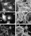
Effects of Rnd1 expression on actin filament assembly. Serum-starved confluent Swiss 3T3 fibroblasts were injected with a pRK5 vector expressing Rnd1 (A–F) or myc-tagged Rnd3/ RhoE (G and H). 1.5–2.5 h later, cells were either left unstimulated for a further 30 min (A and B), stimulated with LPA (100 ng/ml) for 30 min (C, D, G, and H), or stimulated with PDGF (5 ng/ml) for 20 min (E and F) before fixation. Permeabilized cells were stained to show Rnd1 expression (B, D, and F), myc-tagged Rnd3/RhoE expression (H), and actin filaments (A, C, E, and G). Bar, 50 μm.
Figure 8
Figure 8
Effects of mutant Rnd1 expression on actin filament assembly. Serum-starved, confluent Swiss 3T3 fibroblasts were injected with pRK5 vectors expressing N27Rnd1 (A and B) or NH2-terminal truncated Rnd1 (C and D) and 2–3 h later, cells were stimulated with LPA (100 ng/ml) for 30 min before fixation. Permeabilized cells were stained to show Rnd1 expression (A and C) and actin filaments (B and D). Growing primary REFs were microinjected with a pRK5 vector expressing wild-type Rnd1 and fixed 2 h later. Permeabilized cells were costained to show vinculin localization (E) and actin filaments (F). Cells were coinjected with dextran-biotin (lysinated) and visualized with Cascade blue–conjugated avidin. At least 90% of injected cells expressed Rnd1 and showed loss of actin stress fibers and focal adhesions. The REF cell microinjected with Rnd1 is indicated by arrows in E and F. Bar, 50 μm.
Figure 9
Figure 9
Effects of Rnd1 expression on actin filaments in epithelial cells. Confocal analysis of MDCK cells microinjected with a pRK5 vector expressing wild-type Rnd1. Cells were prepermeabilized, fixed 3 h after injection, and then stained to show Rnd1 expression (B and D) and actin filaments (A and C). Images A and B show a confocal section through a region juxtaposed to the basal membrane, and images C and D represent a section through the adherens junction lateral membranes. We are unable to assess whether there is any significance to the nuclear staining pattern of overexpressed Rnd1. It has often been observed that overexpression of other small G proteins leads to nuclear localization of the posttranslationally unprocessed protein.

Similar articles

Cited by

References

    1. Aberle H, Bierkamp C, Torchard D, Serova O, Wagner T, Natt E, Wirshing J, Heidkamper C, Montagna M, Lynch HT, et al. The human plakoglobin gene localizes on chromosome 17 q21 and is subjected to loss of heterozygosity in breast and ovarian cancers. Proc Natl Acad Sci USA. 1995;92:6384–6388. - PMC - PubMed
    1. Aktories K, Just I. In vitro ADP-ribosylation of Rho by bacterial ADP-ribosyl transferases. Methods Enzymol. 1995;256:184–195. - PubMed
    1. Amano M, Ito M, Kimura K, Fukata Y, Chihara K, Nakano T, Matsuura Y, Kaibuchi K. Phosphorylation and activation of myosin by Rho-associated kinase (Rho-kinase) J Biol Chem. 1996;271:20246–20249. - PubMed
    1. Bar-Sagi D, Feramisco JR. Induction of membrane ruffling and fluid-phase pinocytosis in quiescent fibroblasts by Ras proteins. Science. 1986;233:1061–1068. - PubMed
    1. Cerione RA, Zheng Y. The Dbl family of oncogenes. Curr Opin Cell Biol. 1996;8:216–222. - PubMed

Publication types

MeSH terms