iBet uBet web content aggregator. Adding the entire web to your favor.


Link to original content: https://pubmed.ncbi.nlm.nih.gov/9314561
p46, a novel natural killer cell-specific surface molecule that mediates cell activation - PubMed Skip to main page content
U.S. flag

An official website of the United States government

Dot gov

The .gov means it’s official.
Federal government websites often end in .gov or .mil. Before sharing sensitive information, make sure you’re on a federal government site.

Https

The site is secure.
The https:// ensures that you are connecting to the official website and that any information you provide is encrypted and transmitted securely.

Access keys NCBI Homepage MyNCBI Homepage Main Content Main Navigation
. 1997 Oct 6;186(7):1129-36.
doi: 10.1084/jem.186.7.1129.

p46, a novel natural killer cell-specific surface molecule that mediates cell activation

Affiliations

p46, a novel natural killer cell-specific surface molecule that mediates cell activation

S Sivori et al. J Exp Med. .

Abstract

Limited information is available on the surface molecules that are involved in natural killer (NK) cell triggering. In this study, we selected the BAB281 monoclonal antibody (mAb) on the basis of its ability to trigger NK-mediated target cell lysis. BAB281 identified a novel NK cell-specific surface molecule of 46 kD (p46) that is expressed by all resting or activated NK cells. Importantly, unlike the NK cell antigens identified so far, the expression of p46 was strictly confined to NK cells. Upon mAb-mediated cross-linking, p46 molecules induced strong cell triggering leading to [Ca2+]i increases, lymphokine production, and cytolytic activity both in resting NK cells and NK cell clones. The p46-mediated induction of Ca2+ increases or triggering of cytolytic activity was downregulated by the simultaneous engagement of inhibitory receptors including p58, p70, and CD94/NKG2A. Both the unique cellular distribution and functional capability of p46 molecules suggest a possible role in the mechanisms of non-major histocompatibility complex-restricted cytolysis mediated by human NK cells.


Figures
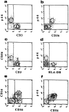
Figure 3
Figure 3
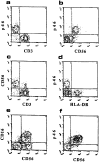
Analysis of the expression of p46 molecules in peripheral blood NK cells. In these experiments freshly isolated PBMCs either unfractionated (a–d) or depleted of both adherent cells and CD3+ lymphocytes (e–g) were analyzed by two-color immunofluorescence and FACS® analysis for the expression of p46 surface molecules in combination with the indicated surface markers. Cells were stained with mAbs to the indicated molecules followed by FITC or PE-conjugated isotype-specific goat anti– mouse second reagent. The contour plots were divided into quadrants representing unstained cells (lower left), cells with only red fluorescence (upper left), cells with red and green fluorescence (upper right), and cells with only green fluorescence (lower right).
Figure 3
Figure 3
Analysis of the expression of p46 molecules in peripheral blood NK cells. In these experiments freshly isolated PBMCs either unfractionated (a–d) or depleted of both adherent cells and CD3+ lymphocytes (e–g) were analyzed by two-color immunofluorescence and FACS® analysis for the expression of p46 surface molecules in combination with the indicated surface markers. Cells were stained with mAbs to the indicated molecules followed by FITC or PE-conjugated isotype-specific goat anti– mouse second reagent. The contour plots were divided into quadrants representing unstained cells (lower left), cells with only red fluorescence (upper left), cells with red and green fluorescence (upper right), and cells with only green fluorescence (lower right).
Figure 6
Figure 6
p46-mediated triggering of cytolytic activity in fresh peripheral blood NK cells. (a) Freshly isolated peripheral blood NK cells were analyzed for their ability to kill the indicated FcγR+ target cells in the absence of mAb (□) or presence of anti-CD16 (▨ ), BAB281 (▪), or anti-CD56 mAb (▤ ). The effector target ratio was 20:1 against P815 target cells and 10:1 against K562 target cells. (b) Freshly isolated PBL were sorted according to the lack of expression of p46 (○) or CD3 (▴) or both p46 and CD3 (*) and compared to unfractionated cells (•) for cytolytic activity against 51Cr-labeled K562 target cells in the absence of added mAbs.
Figure 6
Figure 6
p46-mediated triggering of cytolytic activity in fresh peripheral blood NK cells. (a) Freshly isolated peripheral blood NK cells were analyzed for their ability to kill the indicated FcγR+ target cells in the absence of mAb (□) or presence of anti-CD16 (▨ ), BAB281 (▪), or anti-CD56 mAb (▤ ). The effector target ratio was 20:1 against P815 target cells and 10:1 against K562 target cells. (b) Freshly isolated PBL were sorted according to the lack of expression of p46 (○) or CD3 (▴) or both p46 and CD3 (*) and compared to unfractionated cells (•) for cytolytic activity against 51Cr-labeled K562 target cells in the absence of added mAbs.
Figure 1
Figure 1
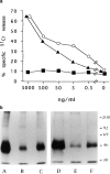
Triggering of cytolytic activity and molecular characteristics of the novel 46-kD NK cell surface molecule. (a) The NK clone SE192, used for mice immunization, was analyzed in a redirected killing assay against P815 target cells in the presence of graded amounts of BAB281 (▴), c127 (anti-CD16) (○), or c218 (anti-CD56) (▪) mAbs. All these mAbs are IgG1. (b) NK cell populations derived from donor E.C. (lanes A and D) or donor L.M. (lanes B and E) and the NK cell clone 211 derived from donor F.G. (lanes C and F) were surface labeled with 125I and immunoprecipitated with BAB281 mAb. Samples were analyzed in a 8% SDS-PAGE under nonreducing (lanes A–C) or reducing (lanes D–F) conditions. All the mAbs used here were of the IgG1 isotype.
Figure 1
Figure 1
Triggering of cytolytic activity and molecular characteristics of the novel 46-kD NK cell surface molecule. (a) The NK clone SE192, used for mice immunization, was analyzed in a redirected killing assay against P815 target cells in the presence of graded amounts of BAB281 (▴), c127 (anti-CD16) (○), or c218 (anti-CD56) (▪) mAbs. All these mAbs are IgG1. (b) NK cell populations derived from donor E.C. (lanes A and D) or donor L.M. (lanes B and E) and the NK cell clone 211 derived from donor F.G. (lanes C and F) were surface labeled with 125I and immunoprecipitated with BAB281 mAb. Samples were analyzed in a 8% SDS-PAGE under nonreducing (lanes A–C) or reducing (lanes D–F) conditions. All the mAbs used here were of the IgG1 isotype.
Figure 2
Figure 2
Deglycosylation and proteolytic digestion of p46 molecules. (a) CD16 (lanes A–D) or p46 (lanes E–H) molecules isolated from a surface labeled NK cell population were treated with neuraminidase (lanes B and F), neuraminidase plus O-glycanase (lanes C and G), or N-glycanase (lanes D and H). Untreated molecules are shown in lanes A and E. Samples were run in an 11% SDS-PAGE under reducing conditions. (b) p46 and CD16 molecules immunoprecipitated from a surface labeled NK cell population were analyzed by SDS-PAGE. Bands corresponding to p46 (lanes A, C, E, and G) or CD16 (lanes B, D, F, and H) molecules were excised from the gel and analyzed by SDS-PAGE on a 15–20% gradient gel under reducing conditions after digestion with V8 protease (lanes C and D) or papain (lanes G and H). In lanes A, B, E, and F untreated molecules are shown.
Figure 2
Figure 2
Deglycosylation and proteolytic digestion of p46 molecules. (a) CD16 (lanes A–D) or p46 (lanes E–H) molecules isolated from a surface labeled NK cell population were treated with neuraminidase (lanes B and F), neuraminidase plus O-glycanase (lanes C and G), or N-glycanase (lanes D and H). Untreated molecules are shown in lanes A and E. Samples were run in an 11% SDS-PAGE under reducing conditions. (b) p46 and CD16 molecules immunoprecipitated from a surface labeled NK cell population were analyzed by SDS-PAGE. Bands corresponding to p46 (lanes A, C, E, and G) or CD16 (lanes B, D, F, and H) molecules were excised from the gel and analyzed by SDS-PAGE on a 15–20% gradient gel under reducing conditions after digestion with V8 protease (lanes C and D) or papain (lanes G and H). In lanes A, B, E, and F untreated molecules are shown.
Figure 4
Figure 4
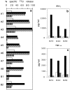
All human NK cell clones can be triggered via p46 molecule. (a) 10 NK cell clones (representative of >100 clones analyzed) were assessed for cytolytic activity against the FcγR+ P815 target cells in the absence (□) or presence of anti-p46 mAb (▪), anti-CD16 mAb (▨ ), or anti-CD56 mAb (▥ ). Two clones (B1 and B2) representative of the infrequent CD56+16 NK cell clones, were triggered only by anti-p46 mAb. (b and c) Three representative NK cell clones were analyzed for IFN-γ or TNF-α production in the absence (□) or presence of anti-p46 mAb (▪) or anti-CD56 mAb (▥ ).
Figure 5
Figure 5
The p46-mediated NK cell triggering is downregulated by the cross-linking of HLA–class I specific inhibitory receptors. Three representative NK cell clones expressing different HLA–class I specific inhibitory receptors were analyzed for cytolytic activity in a redirected killing assay against the FcγR+ P815 target cells in the absence (□) or presence of anti-NKR (▪), anti-p46 (▤ ), both anti-p46 and anti-NKR (▥ ), or both anti-p46 and anti-CD56 (▨ ) mAbs. The anti-NKR mAbs were represented by: EB6 (anti-p58.1) in clone KK26; Z199 (anti-CD94/ NKG2A complex) in clone KK4; and Z27 (anti-p70) in clone LO1.
Figure 7
Figure 7
Anti–p46-mediated [Ca2+]i mobilization. Clone KK26 (p46+, p58.1+, p50.3+) was analyzed for [Ca2+]i mobilization after stimulation with anti-p46 (a) or anti-p46 plus anti-p58.1 (b) mAb followed by isotype-specific goat anti–mouse. (c and d) It is also shown that the [Ca2+]i mobilization induced by anti-p50.3 mAb (c) can be downregulated by the co–cross-linking of the activating molecules (p50.3) with the inhibitory NK receptor (p58.1) (d). All the mAbs used here were of the IgG1 isotype.

Similar articles

Cited by

References

    1. Herberman RB, Nunn ME, Lavrin DH. Natural cytotoxic reactivity of mouse lymphoid cells against syngeneic and allogeneic tumours. I. Distribution of reactivity and specificity. Int J Cancer. 1976;16:216–229. - PubMed
    1. Trinchieri G. Biology of natural killer cells. Adv Immunol. 1989;47:187–376. - PMC - PubMed
    1. Liao NS, Bix M, Zijstra M, Jaenisch R, Raulet D. MHC class I deficiency: susceptibility to natural killer (NK) cells and impaired NK activity. Science (Wash DC) 1991;253:199–202. - PubMed
    1. Karre K. An unexpected petition for pardon. Curr Biol. 1992;2:613–616. - PubMed
    1. Moretta L, Ciccone E, Mingari MC, Biassoni R, Moretta A. Human NK cells: origin, clonality, specificity and receptors. Adv Immunol. 1994;55:341–380. - PubMed

MeSH terms

Substances