The use of dose surface maps as a tool to investigate spatial dose delivery accuracy for the rectum during prostate radiotherapy
- PMID: 38425148
- PMCID: PMC11244681
- DOI: 10.1002/acm2.14314
The use of dose surface maps as a tool to investigate spatial dose delivery accuracy for the rectum during prostate radiotherapy
Abstract
Purpose: This study aims to address the lack of spatial dose comparisons of planned and delivered rectal doses during prostate radiotherapy by using dose-surface maps (DSMs) to analyze dose delivery accuracy and comparing these results to those derived using DVHs.
Methods: Two independent cohorts were used in this study: twenty patients treated with 36.25 Gy in five fractions (SBRT) and 20 treated with 60 Gy in 20 fractions (IMRT). Daily delivered rectum doses for each patient were retrospectively calculated using daily CBCT images. For each cohort, planned and average-delivered DVHs were generated and compared, as were planned and accumulated DSMs. Permutation testing was used to identify DVH metrics and DSM regions where significant dose differences occurred. Changes in rectal volume and position between planning and delivery were also evaluated to determine possible correlation to dosimetric changes.
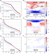
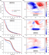
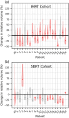
Results: For both cohorts, DVHs and DSMs reported conflicting findings on how planned and delivered rectum doses differed from each other. DVH analysis determined average-delivered DVHs were on average 7.1% ± 7.6% (p ≤ 0.002) and 5.0 ± 7.4% (p ≤ 0.021) higher than planned for the IMRT and SBRT cohorts, respectively. Meanwhile, DSM analysis found average delivered posterior rectal wall dose was 3.8 ± 0.6 Gy (p = 0.014) lower than planned in the IMRT cohort and no significant dose differences in the SBRT cohort. Observed dose differences were moderately correlated with anterior-posterior rectal wall motion, as well as PTV superior-inferior motion in the IMRT cohort. Evidence of both these relationships were discernable in DSMs.
Conclusion: DSMs enabled spatial investigations of planned and delivered doses can uncover associations with interfraction motion that are otherwise masked in DVHs. Investigations of dose delivery accuracy in radiotherapy may benefit from using DSMs over DVHs for certain organs such as the rectum.
Keywords: SBRT; dose delivery; dose reconstruction; prostate; radiotherapy.
© 2024 The Authors. Journal of Applied Clinical Medical Physics is published by Wiley Periodicals, Inc. on behalf of The American Association of Physicists in Medicine.
Conflict of interest statement
No conflicts of interest.
Figures


Similar articles
-
Dosimetric impact of organ at risk daily variation during prostate stereotactic ablative radiotherapy.Br J Radiol. 2020 Apr;93(1108):20190789. doi: 10.1259/bjr.20190789. Epub 2020 Jan 30. Br J Radiol. 2020. PMID: 31971829 Free PMC article.
-
Dosimetric and volumetric changes in the rectum and bladder in patients receiving CBCT-guided prostate IMRT: analysis based on daily CBCT dose calculation.J Appl Clin Med Phys. 2016 Nov 8;17(6):107-117. doi: 10.1120/jacmp.v17i6.6207. J Appl Clin Med Phys. 2016. PMID: 27929486 Free PMC article.
-
Associations between volume changes and spatial dose metrics for the urinary bladder during local versus pelvic irradiation for prostate cancer.Acta Oncol. 2017 Jun;56(6):884-890. doi: 10.1080/0284186X.2017.1312014. Epub 2017 Apr 12. Acta Oncol. 2017. PMID: 28401808 Free PMC article.
-
Accumulated dose to the rectum, measured using dose-volume histograms and dose-surface maps, is different from planned dose in all patients treated with radiotherapy for prostate cancer.Br J Radiol. 2015 Oct;88(1054):20150243. doi: 10.1259/bjr.20150243. Epub 2015 Jul 24. Br J Radiol. 2015. PMID: 26204919 Free PMC article.
-
Delivered dose can be a better predictor of rectal toxicity than planned dose in prostate radiotherapy.Radiother Oncol. 2017 Jun;123(3):466-471. doi: 10.1016/j.radonc.2017.04.008. Epub 2017 Apr 28. Radiother Oncol. 2017. PMID: 28460825 Free PMC article.
References
-
- De Crevoisier R, Tucker SL, Dong L, et al. Increased risk of biochemical and local failure in patients with distended rectum on the planning CT for prostate cancer radiotherapy. Int J Radiat Oncol Biol Phys. 2005;62(4):965‐973. - PubMed
-
- Zelefsky MJ, Kollmeier M, Cox B, et al. Improved clinical outcomes with high‐dose image guided radiotherapy compared with non‐IGRT for the treatment of clinically localized prostate cancer. Int J Radiat Oncol Biol Phys. 2012;84(1):125‐129. - PubMed
-
- Haaren PM, Bel A, Hofman P, Vulpen M, Kotte AN, Heide UA. Influence of daily setup measurements and corrections on the estimated delivered dose during IMRT treatment of prostate cancer patients. Radiother Oncol. 2009;90(3):291‐298. - PubMed
-
- Fraser DJ, Chen Y, Poon E, Cury FL, Falco T, Verhaegen F. Dosimetric consequences of misalignment and realignment in prostate 3DCRT using intramodality ultrasound image guidance. Medical Physics. 2010. - PubMed
MeSH terms
LinkOut - more resources
Full Text Sources
Medical


