Molecular assembly indices of mineral heteropolyanions: some abiotic molecules are as complex as large biomolecules
- PMID: 38378136
- PMCID: PMC10878807
- DOI: 10.1098/rsif.2023.0632
Molecular assembly indices of mineral heteropolyanions: some abiotic molecules are as complex as large biomolecules
Abstract
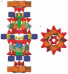
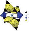
Molecular assembly indices, which measure the number of unique sequential steps theoretically required to construct a three-dimensional molecule from its constituent atomic bonds, have been proposed as potential biosignatures. A central hypothesis of assembly theory is that any molecule with an assembly index ≥15 found in significant local concentrations represents an unambiguous sign of life. We show that abiotic molecule-like heteropolyanions, which assemble in aqueous solution as precursors to some mineral crystals, range in molecular assembly indices from 2 for H2CO3 or Si(OH)4 groups to as large as 21 for the most complex known molecule-like subunits in the rare minerals ewingite and ilmajokite. Therefore, values of molecular assembly indices ≥15 do not represent unambiguous biosignatures.
Keywords: Titan; assembly theory; heteropolyanion; mineral evolution; molecular complexity.
Conflict of interest statement
The authors declare no competing interests.
Figures


Similar articles
-
Planning Implications Related to Sterilization-Sensitive Science Investigations Associated with Mars Sample Return (MSR).Astrobiology. 2022 Jun;22(S1):S112-S164. doi: 10.1089/AST.2021.0113. Epub 2022 May 19. Astrobiology. 2022. PMID: 34904892
-
Nano-atomic scale hydrophobic/philic confinement of peptides on mineral surfaces by cross-correlated SPM and quantum mechanical DFT analysis.J Microsc. 2020 Dec;280(3):204-221. doi: 10.1111/jmi.12923. Epub 2020 Jun 8. J Microsc. 2020. PMID: 32458447
-
Abiotic phosphorus recycling from adsorbed ribonucleotides on a ferrihydrite-type mineral: Probing solution and surface species.J Colloid Interface Sci. 2019 Jul 1;547:171-182. doi: 10.1016/j.jcis.2019.03.086. Epub 2019 Mar 26. J Colloid Interface Sci. 2019. PMID: 30954001
-
Mineral-Lipid Interactions in the Origins of Life.Trends Biochem Sci. 2019 Apr;44(4):331-341. doi: 10.1016/j.tibs.2018.11.009. Epub 2018 Dec 21. Trends Biochem Sci. 2019. PMID: 30583961 Review.
-
The influence of surface active molecules on the crystallization of biominerals in solution.Adv Colloid Interface Sci. 2006 Dec 21;128-130:135-58. doi: 10.1016/j.cis.2006.11.022. Epub 2007 Jan 23. Adv Colloid Interface Sci. 2006. PMID: 17254533 Review.
Cited by
-
Reply to 'Experimental measurement of assembly indices are required to determine the threshold for life'.J R Soc Interface. 2024 Nov;21(220):20240622. doi: 10.1098/rsif.2024.0622. Epub 2024 Nov 20. J R Soc Interface. 2024. PMID: 39563497 Free PMC article.
-
Experimentally measured assembly indices are required to determine the threshold for life.J R Soc Interface. 2024 Nov;21(220):20240367. doi: 10.1098/rsif.2024.0367. Epub 2024 Nov 20. J R Soc Interface. 2024. PMID: 39563496 Free PMC article.
-
Understanding the Role of Layered Minerals in the Emergence and Preservation of Proto-Proteins and Detection of Traces of Early Life.Acc Chem Res. 2024 Sep 3;57(17):2453-2463. doi: 10.1021/acs.accounts.4c00173. Epub 2024 Aug 14. Acc Chem Res. 2024. PMID: 39141709 Free PMC article.
-
On the salient limitations of the methods of assembly theory and their classification of molecular biosignatures.NPJ Syst Biol Appl. 2024 Aug 7;10(1):82. doi: 10.1038/s41540-024-00403-y. NPJ Syst Biol Appl. 2024. PMID: 39112510 Free PMC article. Review.
References
-
- Marshall SM, Moore DG, Murray ARG, Walker SI, Cronin L. 2019. Quantifying the pathways to life using assembly spaces. (https://arxiv.org/pdf/1907.04649.pdf)
-
- Mathis C, Patarroyo KY, Cronin L. 2021. Understanding assembly indices. See http://www.molecular-assembly.com/learn/.
Publication types
MeSH terms
Substances
LinkOut - more resources
Full Text Sources
Medical


