iBet uBet web content aggregator. Adding the entire web to your favor.


Link to original content: https://pubmed.ncbi.nlm.nih.gov/35625743
Osteocalcin Alleviates Lipopolysaccharide-Induced Acute Inflammation via Activation of GPR37 in Macrophages - PubMed Skip to main page content
U.S. flag

An official website of the United States government

Dot gov

The .gov means it’s official.
Federal government websites often end in .gov or .mil. Before sharing sensitive information, make sure you’re on a federal government site.

Https

The site is secure.
The https:// ensures that you are connecting to the official website and that any information you provide is encrypted and transmitted securely.

Access keys NCBI Homepage MyNCBI Homepage Main Content Main Navigation
. 2022 Apr 27;10(5):1006.
doi: 10.3390/biomedicines10051006.

Osteocalcin Alleviates Lipopolysaccharide-Induced Acute Inflammation via Activation of GPR37 in Macrophages

Affiliations

Osteocalcin Alleviates Lipopolysaccharide-Induced Acute Inflammation via Activation of GPR37 in Macrophages

Zhengjiang Qian et al. Biomedicines. .

Abstract

The G protein-coupled receptor 37 (GPR37) has been reported to be expressed in macrophages and the activation of GPR37 by its ligand/agonist, and it can regulate macrophage-associated functions and inflammatory responses. Since our previous work identified that osteocalcin (OCN) acts as an endogenous ligand for GPR37 and can elicit various intracellular signals by interacting with GPR37, we thus hypothesized that OCN may also play a functional role in macrophage through the activation of GPR37. To verify the hypothesis, we conducted a series of in vivo and in vitro studies in lipopolysaccharide (LPS)-challenged mice and primary cultured macrophages. Our results reveal that the OCN gene deletion (OCN-/-) and wild type (WT) mice showed comparable death rates and inflammatory cytokines productions in response to a lethal dose of LPS exposure. However, the detrimental effects caused by LPS were significantly ameliorated by exogenous OCN treatments in both WT and OCN-/- mice. Notably, the protective effects of OCN were absent in GPR37-/- mice. In coordination with the in vivo results, our in vitro studies further illustrated that OCN triggered intracellular responses via GPR37 in peritoneal macrophages by regulating the release of inflammatory factors and macrophage phagocytic function. Finally, we exhibited that the adoptive transfer of OCN-treated macrophages from WT mice significantly inhibits the release of pro-inflammatory cytokines in GPR37-/- mice exposed to LPS. Taken together, these findings suggest a protective role of OCN against LPS-caused acute inflammation, by the activation of GPR37 in macrophages, and provide a potential application of the activation of the OCN/GPR37 regulatory axis as a therapeutic strategy for inflammatory diseases.

Keywords: GPR37; acute inflammation; lipopolysaccharide; macrophage; osteocalcin.


Conflict of interest statement

The authors declare no conflict of interest.

Figures

Figure 1
Figure 1
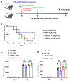
OCN attenuates LPS-caused detrimental effects in WT and OCN−/− mice. (A) Experimental design for the LPS-induced acute inflammation. The OCN−/− and WT mice were injected intraperitoneally with PBS or OCN (500 ng) followed by LPS (10 mg/kg) treatment. Mice survival rate and serum inflammatory were analyzed in each time point, accordingly. (B) Survival curves of OCN−/− and WT mice treated with PBS or OCN in response to LPS stimulation. Sample sizes are presented in brackets (n = 15). * p < 0.05, as compared to PBS. (C,D) ELISA analysis of serum levels of IL-6 (C) and TNFα (D) in OCN−/− and WT mice with PBS or OCN pretreatment after 6h of LPS administration. Sample sizes are indicated as dots in each column. Data are presented as mean ± SD. * p < 0.05 and ** p < 0.01, as compared to PBS.
Figure 2
Figure 2
OCN inhibits the production of pro-inflammatory factor in macrophage. (A,B) Effects of OCN treatment on the mRNA expression of pro-inflammatory genes, i.e., IL-6 (A) and TNFα (B), in LPS (500 ng/mL) treated peritoneal macrophages from WT mice. Data are presented as means ± SD (n = 3 biological replicates). * p < 0.05 and ** p < 0.01, as compared to PBS. (C,D) Cytokine levels in a culture medium of peritoneal macrophage from WT and OCN−/− mice. Peritoneal macrophages were treated with LPS (500 ng/mL), together PBS or OCN (10 nM) for 12 h, and the levels of IL-6 (C) and TNFα (D) in culture medium were measured by ELISA analysis. Data are presented as means ± SD. (n = 3 biological replicates) * p < 0.05, as compared to PBS. (E) The mRNA expression analysis of genes associated with anti-inflammatory in peritoneal macrophages isolated from WT mice. Data are presented as means ± SD (n = 3 biological replicates). ** p < 0.01 and *** p < 0.001, as compared to PBS.
Figure 3
Figure 3
GPR37 mediates OCN-induced intracellular responses in macrophages. (A) Overview of the experimental design testing intracellular signals in macrophages. Peritoneal macrophages were isolated from WT and GPR37−/− mice to study the changes of intracellular calcium level (iCa2+), cAMP production, and pERK level in response to OCN treatment. (B) Representative response curves of OCN-triggered iCa2+ changes in macrophages from WT and GPR37−/− mice. (C) OCN-triggered inhibition of cAMP production in macrophages from WT and GPR37−/− mice. Data are presented as mean ± SD (n = 3 biological replicates). * p < 0.05, as compared to PBS, and n.s. indicates not significant. (D,E) Representative immunoblot images (D) and relative quantification (E) showing that OCN-triggered an increase in the pERK level in macrophages from WT but not in that from GPR37−/− mice. Data are presented as mean ± SD (n = 3 biological replicates). * p < 0.05, as compared to PBS. (F) Representative response curves of OCN-triggered iCa2+ changes in PTX pretreated macrophage from WT mice. (G) OCN-triggered inhibition of cAMP production in PTX pretreated macrophage from WT mice. Data are presented as mean ± SD (n = 3 biological replicates). * p < 0.05, as compared to PBS, and n.s. indicates not significant. (H,I) Representative immunoblot images (H) and relative quantification (I) showing that PTX pretreatment blocked the OCN-triggered increase in the pERK level in macrophages from WT mice. Data are presented as mean ± SD (n = 3 biological replicates). * p < 0.05, as compared to PBS.
Figure 4
Figure 4
GPR37 mediates the inhibitory effects of OCN on pro-inflammatory factors in macrophage. (A,B) Cytokine levels in the culture medium of peritoneal macrophages from WT and GPR37−/− mice. Cells were treated with LPS (500 ng/mL, 37 °C, 24 h) together with PBS or OCN (10 nM). ELISA analysis was performed to measure the level of IL-6 (A) and TNF-α (B). Data are presented as mean ± SD (n = 3 biological replicates), * p < 0.05, as compared to PBS, and n.s. indicates not significant. (C,D) Representative immunoblot images (C) and quantitative analysis (D) of NFκB p65 level in macrophages treated with LPS in the presence of OCN. Data are presented as mean ± SD (n = 3 biological replicates), * p < 0.05 as compared to PBS, and n.s. indicates not significant.
Figure 5
Figure 5
OCN promotes macrophage phagocytosis via GPR37. (A) Representative images of in vitro phagocytosis assay, in which pHrodo zymosan particles were incubated with peritoneal macrophages from WT or OCN−/− mice, treated with PBS or OCN (10 nM, 30 min, 37 °C). Red fluorescence suggests an intracellular update indicating phagocytosis. Scale bars: 20 μm. (B) Quantification of macrophage phagocytic activity of zymosan positives cells. Data are presented as mean ± SD (n = 3 biological replicates), ** p < 0.01, as compared to PBS. (C) Representative images of an in vitro phagocytosis assay, in which pHrodo zymosan particles were incubated with peritoneal macrophages from WT or GPR37−/− mice, treated with PBS or OCN (10 nM, 30 min, 37 °C). Scale bars: 20 μm. (D) Quantification of macrophage phagocytic activity of zymosan positives cells. Data are presented as mean ± SD (n = 3 biological replicates), ** p < 0.01, as compared to PBS, and n.s. indicates not significant.
Figure 6
Figure 6
The protective function of OCN against LPS is absent in GPR37−/− mice. (A) Experimental design for LPS-induced acute inflammation in GPR37−/− and WT mice. After intraperitoneal injection of PBS or OCN (500 ng), followed by LPS (10 mg/kg) treatment, mice survival rate and serum inflammatory cytokine level were analyzed accordingly. (B) Survival curves of GPR37−/− and WT mice treated with PBS or OCN in response to an LPS challenge. Sample sizes are presented in brackets (n = 15). * p < 0.05, as compared to PBS. (C,D) After 6h of LPS injection, with or without OCN pretreatment, serum IL-6 (C) and TNFα (D) levels were analyzed in GPR37−/− and WT mice. Sample sizes are indicated as dots in columns. * p < 0.05 and ** p < 0.01, as compared to PBS; n.s. indicates not significant.
Figure 7
Figure 7
Adoptive transfer of OCN-treated macrophages confers protection against LPS stimulation. (A) Experimental design, to test whether adoptive transfer of macrophages pretreated with OCN, could attenuate LPS-induced acute inflammation. Peritoneal macrophages were isolated from WT or GPR37−/− mice and then treated with PBS or OCN (10 nM) for 24 h, followed by a washout of OCN and an adoptive transfer of macrophages (I.P.) into GPR37−/− mice challenged with LPS for 1 h. (B,C) ELISA analysis of serum IL-6 (B) and TNFα (C) levels, in GPR37−/− mice with adoptive transfer, of macrophages derived from WT or GPR37−/− mice. Sample sizes are indicated as dots in each column. Data are presented as mean ± SD. * p < 0.05 and ** p < 0.01, as compared to PBS; n.s. indicates not significant.
Figure 8
Figure 8
A schematic diagram illustrating that the bone-derived hormone OCN, via the activation of GPR37 in macrophages, plays a protective function against LPS challenge through the regulation of macrophage inflammatory reactions and phagocytic function.

Similar articles

Cited by

References

    1. Hauschka P.V., Lian J.B., Cole D.E., Gundberg C.M. Osteocalcin and matrix Gla protein: Vitamin K-dependent proteins in bone. Physiol. Rev. 1989;69:990–1047. doi: 10.1152/physrev.1989.69.3.990. - DOI - PubMed
    1. Li J., Zhang H., Yang C., Li Y., Dai Z. An overview of osteocalcin progress. J. Bone Miner. Metab. 2016;34:367–379. doi: 10.1007/s00774-015-0734-7. - DOI - PubMed
    1. Komori T. What is the function of osteocalcin? J. Oral Biosci. 2020;62:223–227. doi: 10.1016/j.job.2020.05.004. - DOI - PubMed
    1. Schatz M., Saravanan S., d’Adesky N.D., Bramlett H., Perez-Pinzon M.A., Raval A.P. Osteocalcin, ovarian senescence, and brain health. Front. Neuroendocrinol. 2020;59:100861. doi: 10.1016/j.yfrne.2020.100861. - DOI - PubMed
    1. Moser S.C., van der Eerden B.C.J. Osteocalcin-A Versatile Bone-Derived Hormone. Front. Endocrinol. 2018;9:794. doi: 10.3389/fendo.2018.00794. - DOI - PMC - PubMed

LinkOut - more resources