iBet uBet web content aggregator. Adding the entire web to your favor.


Link to original content: https://pubmed.ncbi.nlm.nih.gov/35234892
The RecD2 helicase balances RecA activities - PubMed Skip to main page content
U.S. flag

An official website of the United States government

Dot gov

The .gov means it’s official.
Federal government websites often end in .gov or .mil. Before sharing sensitive information, make sure you’re on a federal government site.

Https

The site is secure.
The https:// ensures that you are connecting to the official website and that any information you provide is encrypted and transmitted securely.

Access keys NCBI Homepage MyNCBI Homepage Main Content Main Navigation
. 2022 Apr 8;50(6):3432-3444.
doi: 10.1093/nar/gkac131.

The RecD2 helicase balances RecA activities

Affiliations

The RecD2 helicase balances RecA activities

Cristina Ramos et al. Nucleic Acids Res. .

Abstract

DNA helicases of the RecD2 family are ubiquitous. Bacillus subtilis RecD2 in association with the single-stranded binding protein SsbA may contribute to replication fork progression, but its detailed action remains unknown. In this work, we explore the role of RecD2 during DNA replication and its interaction with the RecA recombinase. RecD2 inhibits replication restart, but this effect is not observed in the absence of SsbA. RecD2 slightly affects replication elongation. RecA inhibits leading and lagging strand synthesis, and RecD2, which physically interacts with RecA, counteracts this negative effect. In vivo results show that recD2 inactivation promotes RecA-ssDNA accumulation at low mitomycin C levels, and that RecA threads persist for a longer time after induction of DNA damage. In vitro, RecD2 modulates RecA-mediated DNA strand-exchange and catalyzes branch migration. These findings contribute to our understanding of how RecD2 may contribute to overcome a replicative stress, removing RecA from the ssDNA and, thus, it may act as a negative modulator of RecA filament growth.


Figures

Figure 1.
Figure 1.
RecD2 action on replication restart. (A) RecD2 interaction with SsbA and RecA is detected by immuno-dot blot. Increasing concentrations of RecA, SsbA and BSA (from 100–400 ng) were bound to a nitrocellulose membrane, which was subsequently incubated with a buffer containing 200 ng/ml of purified RecD2. After washing, the membrane was incubated with a mouse monoclonal anti-His antibody. (B) The mini-circular DNA template mimics a blocked replication fork with a 396-nt gap in the lagging strand. Diagram of the DNA template and B. subtilis replisome assembly scheme. Replisomes were assembled in the presence of RecD2 helicase incubating the minicircle DNA replication template in the presence of 5 μM ATPγS. Reactions were started by the addition of dNTPs and ATP, and stopped after the indicated times. (C) Leading strand synthesis in the presence of increasing amounts RecD2. (D) Lagging strand synthesis in the presence of increasing amounts of RecD2. Replication products were analyzed by alkaline gel electrophoresis and autoradiography. Ld, leading; Lg, lagging; M, molecular weight marker (radiolabeled λ-HindIII DNA). (E) Quantification of leading strand synthesis obtained after 2 min with increasing amounts of RecD2 (0–20 nM). (F) Quantification of lagging strand synthesis obtained after 2 min with increasing amounts of RecD2 (0–20 nM). Results are plotted as the mean ± SD of five independent experiments.
Figure 2.
Figure 2.
RecD2 slightly affects ongoing DNA replication. The B. subtilis replisome was assembled on the DNA template and replication was allowed to start by dNTPs and ATP addition. After 1 min, increasing concentrations of RecD2 were added and replication continued for 5 min. (A) A representative alkaline agarose gel showing leading strand and lagging strand synthesis. Ld, leading; Lg, lagging. M, molecular weight marker. (B) Quantification of DNA synthesis. Represented is the mean of three independent experiments ± SD.
Figure 3.
Figure 3.
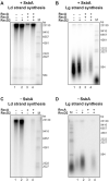
The RecD2 helicase overcomes the inhibition of replication restart by RecA. In A and B: Reactions in the presence of SsbA. An enzyme mix having B. subtilis replisome proteins was added to a substrate mix containing the DNA template, SsbA 90 nM, RecD2 5 nM, RecO 50 nM and RecA 500 nM, and DNA replication was allowed for 2 min at 37°C. (A) Leading strand synthesis. (B) Lagging strand synthesis. In C and D: Reactions without SsbA. An enzyme mix having B. subtilis replisome proteins, but no SsbA was added to a substrate mix having the DNA template, RecD2 5 nM, and RecA 500 nM, and DNA replication was allowed for 2 min at 37°C. (C) Leading strand synthesis. (D) Lagging strand synthesis. In Supplementary Figure S6, the quantifications of DNA synthesis from three independent experiments are shown.
Figure 4.
Figure 4.
RecD2 modulates strand exchange. (A) Strand exchange reactions with RecD2 added at time 0. KpnI-linerarized pGEM-3Zf(+) dsDNA (lds, 3 nM) and its homologous circular ssDNA (css, 3 nM) were incubated with RecA (1.5 μM), RecO (200 nM), SsbA (300 nM) and variable concentrations of RecD2 (1.5 to 50 nM) for 5 min at 37°C with all buffer components except ATP. Then, ATP was added and reactions continued for 20 or 40 min. DNA species were separated by 0.8% w/v agarose gel electrophoresis. RecD2 concentration in lanes 7 and 8: 1.5 nM; lanes 9 and 10: 3 nM, lanes 11 and 12: 6 nM, lanes 13 and 14: 12.5 nM, lanes 15–16: 25 nM, lanes 17–18: 50 nM. In lane 1 a control with the running position of supercoiled and nicked DNA is shown. (B) Quantification of percentage of joint molecules (jm) intermediates and nicked products (nc) after 20 min. Results are the mean of three independent experiments ± SD. (C) Quantification of jm and nc after 40 min. Results are the mean of three independent experiments ± SD. (D) Strand exchange reactions with RecD2 added after strand exchange intitiates at 20 min. lds and css were incubated with RecA, RecO, and SsbA for 20 min at 37°C with all buffer components. Then, variable concentrations of RecD2 (3 to 50 nM) were added and reactions continued for another 20 min (total time 40 min). Lane 3 is a control of the extent of the strand exchange reaction when RecD2 is added (i.e. 20 min reaction). In lanes 4 and 5, two controls of 40 min reactions without RecD2. (E) Quantification of jm and nc observed when RecD2 is added after strand-exchange has been initiated. Plotted are the results from three independent experiments ± SD.
Figure 5.
Figure 5.
RecD2 branch migrates the recombination intermediate. (A) Scheme of the assay. Six standard strand exchange reactions were performed for 10 min at 37°C and pooled. After deproteinization, DNA was purified by gel filtration and ethanol precipitation. The DNA pellet was resuspended in MilliQ water and this DNA, that contains remaining substrates (lds and css) and recombination intermediates (jm), was used to test RecD2 branch migration activity. (B) An assay showing branch migration activity with increasing concentrations of RecD2 (from 3 to 24 nM) in a 30 min reaction. In lane 4, DNA was incubated with all buffer components without RecD2. C3 (lane 3) is a control where the DNA with all buffer components and no protein was kept on ice. Other controls are: C1, plasmid dsDNA (supercoiled, cds and nicked, nc), C2, linear dsDNA (lds) and circular ssDNA (css).
Figure 6.
Figure 6.
Epifluorescence microscopy showing that RecA assembly into threads is affected by RecD2. Quantitative analysis of RecA being diffuse, forming spots or threads in wt (A) or ΔrecD2 (B) cells after treatment with MMC at the indicated times. NT, not treated cells. The results are the average of three independent experiments (n = 400 cells). The corresponding movies are displayed in the Supplementary data (movie S1 and S2, respectively).
Figure 7.
Figure 7.
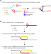
Model depicting how RecD2 helicase may modulate RecA during replication restart and strand exchange. (A) On a stalled fork, RecA may bind to the ssDNA regions inhibiting replication restart. RecD2 removes RecA from the stalled fork and thereby, facilitates replisome loading and replication restart. (B) During homologous recombination the invading strand is coated with RecA. RecD2 may translocate 5′→3′ along the ssDNA and remove RecA. However, once RecA initiates strand exchange it may accelerate heteroduplex extension.

Similar articles

Cited by

References

    1. Lopez de Saro F.J. Regulation of interactions with sliding clamps during DNA replication and repair. Curr. Genomics. 2009; 10:206–215. - PMC - PubMed
    1. Costes A., Lecointe F., McGovern S., Quevillon-Cheruel S., Polard P.. The C-terminal domain of the bacterial SSB protein acts as a DNA maintenance hub at active chromosome replication forks. PLoS Genet. 2010; 6:e1001238. - PMC - PubMed
    1. Shereda R.D., Kozlov A.G., Lohman T.M., Cox M.M., Keck J.L.. SSB as an organizer/mobilizer of genome maintenance complexes. Crit. Rev. Biochem. Mol. Biol. 2008; 43:289–318. - PMC - PubMed
    1. Kreuzer K.N., Brister J.R.. Initiation of bacteriophage T4 DNA replication and replication fork dynamics: a review in the Virology Journal series on bacteriophage T4 and its relatives. Virol. J. 2010; 7:358. - PMC - PubMed
    1. Lo Piano A., Martinez-Jimenez M.I., Zecchi L., Ayora S.. Recombination-dependent concatemeric viral DNA replication. Virus Res. 2011; 160:1–14. - PubMed

Publication types

MeSH terms