iBet uBet web content aggregator. Adding the entire web to your favor.


Link to original content: https://pubmed.ncbi.nlm.nih.gov/35114688
ACE2 binding is an ancestral and evolvable trait of sarbecoviruses - PubMed Skip to main page content
U.S. flag

An official website of the United States government

Dot gov

The .gov means it’s official.
Federal government websites often end in .gov or .mil. Before sharing sensitive information, make sure you’re on a federal government site.

Https

The site is secure.
The https:// ensures that you are connecting to the official website and that any information you provide is encrypted and transmitted securely.

Access keys NCBI Homepage MyNCBI Homepage Main Content Main Navigation
. 2022 Mar;603(7903):913-918.
doi: 10.1038/s41586-022-04464-z. Epub 2022 Feb 3.

ACE2 binding is an ancestral and evolvable trait of sarbecoviruses

Affiliations

ACE2 binding is an ancestral and evolvable trait of sarbecoviruses

Tyler N Starr et al. Nature. 2022 Mar.

Abstract

Two different sarbecoviruses have caused major human outbreaks in the past two decades1,2. Both of these sarbecoviruses, SARS-CoV-1 and SARS-CoV-2, engage ACE2 through the spike receptor-binding domain2-6. However, binding to ACE2 orthologues of humans, bats and other species has been observed only sporadically among the broader diversity of bat sarbecoviruses7-11. Here we use high-throughput assays12 to trace the evolutionary history of ACE2 binding across a diverse range of sarbecoviruses and ACE2 orthologues. We find that ACE2 binding is an ancestral trait of sarbecovirus receptor-binding domains that has subsequently been lost in some clades. Furthermore, we reveal that bat sarbecoviruses from outside Asia can bind to ACE2. Moreover, ACE2 binding is highly evolvable-for many sarbecovirus receptor-binding domains, there are single amino-acid mutations that enable binding to new ACE2 orthologues. However, the effects of individual mutations can differ considerably between viruses, as shown by the N501Y mutation, which enhances the human ACE2-binding affinity of several SARS-CoV-2 variants of concern12 but substantially decreases it for SARS-CoV-1. Our results point to the deep ancestral origin and evolutionary plasticity of ACE2 binding, broadening the range of sarbecoviruses that should be considered to have spillover potential.


Conflict of interest statement

J.D.B. consults for Moderna on viral evolution and epidemiology and Flagship Labs 77 on deep mutational scanning. J.D.B. may receive a share of IP revenue as an inventor on a Fred Hutchinson Cancer Research Center-optioned technology/patent (US Patent and Trademark Office application WO2020006494) related to deep mutational scanning of viral proteins. The Veesler laboratory (S.K.Z., A.C.W. and D.V.) has received an unrelated sponsored research agreement from Vir Biotechnology.

Figures

Fig. 1
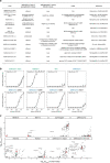
Fig. 1. High-throughput survey of sarbecovirus ACE2 binding.
a, Maximum likelihood phylogeny of sarbecovirus RBDs constructed from RBD nucleotide sequences. The node labels indicate bootstrap support values. Details on rooting are shown in Extended Data Fig. 1. Scale bar, 0.5 nucleotide substitutions per site. b, Binding avidities of sarbecovirus RBDs for eight ACE2 orthologues determined using high-throughput yeast-displayed RBD titration assays (Extended Data Fig. 2). c, Alignment of tested ACE2 orthologues within RBD-contact positions (4 Å cut-off in Protein Data Bank (PDB) 6M0J or 2AJF). d, Representative ACE2-binding curves from high-throughput titrations. Underlying titration curves for individual replicate-barcoded representatives of a genotype are shown in light grey, and the average binding across all barcodes is indicated in black. e, BLI binding analysis of 1 µM R. affinis ACE2–Fc binding to biotinylated BtKY72 RBD immobilized at the surface of streptavidin biosensors (see Extended Data Fig. 3a for analysis of the robustness of the result to ACE2–Fc concentration). Data are representative of three assays using independent preparations of RBD (biological triplicate). f, Entry of VSV particles pseudotyped with the BtKY72 spike into HEK293T cells transiently expressing R. affinis ACE2 alleles 9479 or 787. Each point represents the mean of technical triplicates for assays performed with independent preparation of pseudoviral particles (biological duplicate). The geometric mean is shown by the horizontal line. The normalized pseudovirus western blot, and mock (VSV prepared without spike plasmid) pseudovirus entry in R. affinis ACE2 HEK293T cells are shown in Extended Data Fig. 3c, d.
Fig. 2
Fig. 2. Ancestral origins of sarbecovirus ACE2 binding.
a, Clade-collapsed RBD phylogeny. The circles represent nodes at which ancestral sequences were inferred. The bars indicate putative gains and losses in ACE2 binding. b, ACE2 binding of ancestrally reconstructed, yeast-displayed RBDs (Extended Data Figs. 5 and 6). c, ACE2 binding of AncAsia RBD plus introduction of the 48 substitutions or 2 sequence deletions that occurred on the phylogenetic branch leading to AncClade2 RBD.
Fig. 3
Fig. 3. Evolutionary plasticity of ACE2 binding.
a, The structural context of positions targeted for mutagenesis. Green cartoon, RBD; grey cartoon, ACE2 interaction motifs; blue spheres, residues targeted through mutagenesis (SARS-CoV-2 identities). b, Mutational scanning measurements. The red bars mark the binding avidity of the parental RBD, and the points mark mutant avidities (see Extended Data Fig. 7 for mutation-level measurements). c, The fraction of the 14 RBD backgrounds for which the parental RBD binds to the indicated ACE2 orthologue (−log10(KD,app) > 7), a single mutant binds but the parental RBD does not, or no tested mutants bind. d, Binding of 1 µM human ACE2–Fc to biotinylated RBDs immobilized at the surface of streptavidin biosensors (see Extended Data Fig. 3b for an analysis of the robustness of the result to ACE2–Fc concentration). Data are representative of three assays using independent preparations of RBD (biological triplicate). e, Entry of BtKY72 spike-pseudotyped VSV in HEK293T cells stably expressing human ACE2. Each point represents the mean of technical triplicates in assays performed with independent preparation of pseudoviral particles (biological triplicate). The horizontal line shows the geometric mean. Mock, VSV particles produced in cells in which no spike gene was transfected. A western blot of pseudotyped particles is shown in Extended Data Fig. 3c, and entry into HEK293T cells lacking ACE2 is shown in Extended Data Fig. 3e. f, Titration curves illustrating the effect of mutation to tyrosine 501 (SARS-CoV-2 numbering) in the SARS-CoV-2 and SARS-CoV-1 Urbani RBD backgrounds. g, Epistatic turnover in mutation effects. Each point represents, for a pair of RBDs, the mean absolute error (residual) in their correlated mutant avidities for human ACE2 (Extended Data Fig. 9a) versus their pairwise amino acid sequence identity. Correlations were computed only for pairs in which the parental RBDs bind with −log10(KD,app) > 7. Data are LOESS mean (blue line) ± 95% confidence intervals trendline (grey shading) (see Extended Data Fig. 9b for an analysis across all ACE2 orthologues).
Fig. 4
Fig. 4. Newly sampled sarbecovirus lineages bind to ACE2.
a, Phylogenetic placement of the newly described sarbecovirus RBDs. The new sequences are shown in bold font. RBDs are coloured according to the key in Fig. 1a (Extended Data Fig. 10). Scale bar, expected nucleotide substitutions per site. bd, Binding curves for newly described sarbecovirus RBDs from Europe (b), Africa (c) and Asia (d), and candidate mutations that confer human ACE2 binding. Measurements were performed with yeast-displayed RBDs and purified dimeric ACE2 proteins, measured using flow cytometry. Data are from a single experimental replicate.
Extended Data Fig. 1
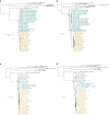
Extended Data Fig. 1. Robustness of the root of the sarbecovirus ingroup.
To establish robustness of our conclusion that the first sarbecovirus divergence is between sarbecoviruses from Africa and Europe and those from Asia, we inferred phylogenies based on alignments of RBD (SARS-CoV-2 spike residues N331-T531) (a,b) or the full spike gene (c,d) and nucleotide (a, c) or amino-acid (b, d) alignments and substitution models. In all four cases, the first sarbecovirus bipartition is placed between sarbecoviruses in Africa/Europe and those in Asia. The placement of the overall tree root is arbitrary with respect to the relationship among non-sarbecovirus outgroups, but this arbitrary placement does not impact the sarbecovirus ingroup rooting. The primary variations among trees includes a potential paraphyletic separation of BtKY72 and BM48–31 from Europe and Africa such that they do not form a monophyletic clade (b; also seen in Extended Data Fig. 10a-c), and variation in the relationships among the three Asia sarbecovirus clades (whose relationship is also inferred with a very low bootstrap support value in our primary phylogeny in Fig. 1a). Known recombination of RBDs with respect to other spike segments among viruses creates incongruencies between spike and RBD trees among Asian sarbecovirus lineages (e.g. ZC45 and ZXC21), though recombination has not been reported among the Africa and Europe spikes and those in Asia. Scale bar, expected number nucleotide or amino-acid substitutions per site. Node labels illustrate bootstrap support values for sarbecovirus and Asia sarbecovirus monophyly. Sequences colored by their RBD clade as in Fig. 1a.
Extended Data Fig. 2
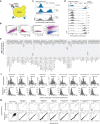
Extended Data Fig. 2. Experimental details of Sort-seq assays.
a, RBD yeast-surface display enables detection of folded RBD expression and ACE2 binding. b, Representative gating for single (SSC-A versus FSC-A, SSC-W versus SSC-H, and FSC-W versus FSC-W), RBD+ (FITC versus FSC-A) cells. c, Representative bins drawn on single cells for expression Sort-seq measurements. d, Representative bins drawn on single, RBD+ cells for ACE2 Tite-seq, measurements. e, Per-variant expression, shown as violin plots across replicate barcodes representing each variant within the gene libraries. f, Number of distinct barcodes for each parental (top) or mutant (bottom) RBD genotype used in the determination of final pooled measurements across libraries. g, Correlation in measured phenotypes between independently assembled and barcoded gene library duplicates for parental (top) or mutant (bottom) RBD genotypes.
Extended Data Fig. 3
Extended Data Fig. 3. Normalization and controls for biolayer interferometry binding and pseudovirus entry assays.
a,b Biolayer interferometry binding analysis of a range of R. affinis ACE2-Fc (a) or human ACE2-Fc (b) concentrations to biotinylated BtKY72 RBD (parental or mutant) immobilized at the surface of streptavidin biosensors. c, Representative Western blots for quantification of spike incorporation into pseudoviral particles. Anti-FLAG (Sigma F3165) identifies incorporation of 3xFLAG-tagged spike, and anti-VSV-M (Kerafast EB0011) identifies level of VSV backbone. Viral inputs into cell entry assays were normalized across pseudoviral particles by S incorporation as determined in the anti-FLAG Western blot. Blot representative of biological duplicate generations of each pseudovirus. For gel source data, see Supplementary Fig. 1. d, Entry into R. affinis ACE2-expressing HEK293T cells by mock VSV particles produced in cells in which no spike gene was transfected. Each point represents the mean of technical triplicates for assays performed with independent preparation of pseudoviral particles (biological replicates). e, Entry of pseudoviral particles into HEK293T cells not transfected with any ACE2. Each point represents the mean of technical triplicates for assays performed with independent preparation of pseudoviral particles (biological replicates).
Extended Data Fig. 4
Extended Data Fig. 4. Clade 2 RBD binding to an expanded panel of R. sinicus ACE2 alleles.
Binding curves for Clade 1a (SARS-CoV-1 Urbani and RsSHC014) and Clade 2 (YN2013 and HKU3-1) sarbecovirus RBDs for 8 R. sinicus ACE2 alleles. Measurements performed with yeast-displayed RBDs and purified dimeric ACE2 proteins, measured by flow cytometry. Data from a single experimental replicate. Region of sampling for bat sarbecovirusess and R. sinicus ACE2 alleles are provided. RsSHC014, YN2013, and HKU3-1 were all sampled from R. sinicus bats.
Extended Data Fig. 5
Extended Data Fig. 5. Full set of RBD ancestral sequence reconstructions.
a, Phylogeny with labelled nodes representing all ancestors tested, including nodes within the SARS-CoV-1 and SARS-CoV-2 clades leading to the human viruses. Branches are annotated with the number of amino-acid substitutions and indels that are inferred to have occurred along each branch. b, Phenotypes of all most plausible ancestral sequences (including repetition of the data represented in Fig. 2b).
Extended Data Fig. 6
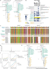
Extended Data Fig. 6. Robustness to uncertainties in ancestral reconstructions.
a, We performed ancestral sequence reconstructions on phylogenies constraining sister relationships between SARS-CoV-2 clade and clade 2 (tree1) or SARS-CoV-1 and SARS-CoV-2 clades (tree2) due to ambiguity in these relationships (Fig. 1a and Extended Data Fig. 1). b, ACE2 binding of alternative reconstructions. “Alt” ancestors incorporate all secondary reconstructed states with posterior probability > 0.2; “tree1” and “tree2” ancestors are inferred on the constrained trees in (a); and “ins117-118” tests the ambiguity of an indel separate from the remaining substitutions in AncSarbecovirus_alt. Sequence differences are listed at right relative to the maximum a posteriori (MAP) ancestors from Fig. 2b and Extended Data Fig. 5b. Mutations are colored red if they were sufficient to abolish the ancestral phenotype and blue if they reinforced it (Extended Data Fig. 7). Dramatic changes to inferred ancestral phenotypes are mostly observed in the alt ancestors which are the most probabilistically distant, while the tree1 and tree2 alternatives generally recapitulate the MAP phenotypes. The exception is AncSARS1a, where the tree1 and tree2 alternatives better match what would be expected based on the descendent RBD phenotypes (Fig. 1b). c, RBD amino acid alignment, indicating a potential recombination breakpoint identified by GARD (from underlying nucleotide sequence). d, Relative support values for possible recombination breakpoints. e, Phylogenies inferred for the putative non-recombinant RBD segments. Arrows point to key changes in the segment 2 sub-tree. Each change is supported by weak bootstrap support values, and this hypothesis introduces a non-parsimonious history with respect to an indel at position 482. We reconstructed AncSarbecovirus_GARD and AncAsia_GARD as concatenated segment 1 and 2 reconstructions. Mutations that distinguish the GARD and MAP ancestor are listed at bottom. f, Binding of GARD ancestors to human and R. affinis 9479 ACE2 was determined in isogenic yeast-display titrations.
Extended Data Fig. 7
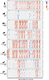
Extended Data Fig. 7. Binding of RBD single mutants to each ACE2.
Each heatmap square illustrates the change in binding caused by the indicated mutation at the indicated position (SARS-CoV-2 numbering), according to the color key shown on the upper-right. Yellow, mutations that were absent from the library or not sampled with sufficient depth in a particular experiment. x markers indicate the wildtype state at each position in each RBD background.
Extended Data Fig. 8
Extended Data Fig. 8. Existing data on sarbecoviruses in mice, and affinities of RBDs and key mutants for mouse versus human ACE2.
a, Summary of infectivity and pathogenesis of natural sarbecovirus and mouse-adapted strains from prior studies,–,–. b, High-throughput titration curves for relevant genotypes from (a). Details as in Fig. 1d. Strength of binding to mouse ACE2 explains the infectivity and pathogenesis of SARS-CoV-1 Urbani and RsSHC014,, relative to the weak or absent replication of WIV1 and SARS-CoV-2 in mice. Mutagenesis data explain the inefficient mouse infectivity of the SARS-CoV-2 B.1.1.7 isolate which incorporates the N501Y RBD mutation, relative to the efficient replication of the mouse-adapted SARS-CoV-2 isolate containing Q498Y or the pathogenic WBP-1 strain containing Q493K and Q498H. c, An ideal mouse-adapted laboratory sarbecovirus strain would bind mouse ACE2 but not human ACE2 due to biosafety considerations. The large red points indicate the affinity of the parental RBD for human and mouse ACE2. The smaller black points indicate mutations, and key mutations that enhance binding to mouse versus human ACE2 are labelled (using SARS-CoV-2 numbering). Further mouse ACE2 specificity may be enabled via mutations at other positions not surveyed in our set of six positions.
Extended Data Fig. 9
Extended Data Fig. 9. Epistasis and turnover in mutational effects.
a, Example correlations in binding affinities for mutants in distinct RBD backgrounds at each site for human ACE2. Plots illustrate mutant avidities for human ACE2 and mean absolute error (residual) in the correlation for mutation measurements in GD-Pangolin (top) and SARS-CoV-1 Urbani (bottom) versus SARS-CoV-2. Plotting symbols indicate amino acid for each measurement. b, Epistatic turnover in mutational effects across RBD backgrounds. Details as in Fig. 3g, but incorporating mutation effects among RBD pairs across all tested ACE2s. Blue line and shaded grey, LOESS mean and 95% CI trendline. See Extended Data Fig. 9b for analysis across all ACE2 orthologues.
Extended Data Fig. 10
Extended Data Fig. 10. Robustness of rooting and AncSarbecovirus phenotype in a phylogeny incorporating newly reported sequences.
a-d, Phylogenetic inference with inclusion of newly reported sarbecovirus sequences (Fig. 4a). As in Extended Data Fig. 1, we infer phylogenies with RBD (a,b) and full spike alignments (c,d), both on nucleotide sequences (a,c) and translated amino acid (b,d) sequence alignments. The full set of outgroup betacoronavirus sequences shown in Extended Data Fig. 1 were also included in this tree inference but truncated from the display for visual clarity. The phylogeny in Fig. 4a is a constrained version of the RBD nucleotide tree from (a) where we constrained a monophyletic relationship among Africa/Europe sarbecoviruses due to uncertainty in the exact placement of the root within or relative ot the Africa/Europe sarbecovirus clade. e, ACE2 binding by parental RBD and candidate mutants in an updated AncSarbecovirus sequence (“v2”) inferred from the phylogeny in Fig. 4a that incorporates many newly described sarbecovirus RBDs, including some in important new phylogenetic locations. The unconstrained tree in (a) leads to inference of an AncSarbecovirus sequence that is identical to Khosta-2 (which also binds ACE2). Sequence differences between the original MAP AncSarbecovirus and the “v2” reconstruction are listed at top. Measurements performed with yeast-displayed RBDs and purified dimeric ACE2 proteins, measured by flow cytometry. Data from a single experimental replicate.

Similar articles

Cited by

References

    1. Cui J, Li F, Shi Z-L. Origin and evolution of pathogenic coronaviruses. Nat. Rev. Microbiol. 2019;17:181–192. - PMC - PubMed
    1. Zhou P, et al. A pneumonia outbreak associated with a new coronavirus of probable bat origin. Nature. 2020;579:270–273. - PMC - PubMed
    1. Li W, et al. Angiotensin-converting enzyme 2 is a functional receptor for the SARS coronavirus. Nature. 2003;426:450–454. doi: 10.1038/nature02145. - DOI - PMC - PubMed
    1. Walls AC, et al. Structure, function, and antigenicity of the SARS-CoV-2 spike glycoprotein. Cell. 2020;181:281–292. - PMC - PubMed
    1. Wrapp D, et al. Cryo-EM structure of the 2019-nCoV spike in the prefusion conformation. Science. 2020;367:1260–1263. - PMC - PubMed

MeSH terms

Substances

Supplementary concepts