iBet uBet web content aggregator. Adding the entire web to your favor.


Link to original content: https://pubmed.ncbi.nlm.nih.gov/34679832
The Tongue in Three Species of Lemurs: Flower and Nectar Feeding Adaptations - PubMed Skip to main page content
U.S. flag

An official website of the United States government

Dot gov

The .gov means it’s official.
Federal government websites often end in .gov or .mil. Before sharing sensitive information, make sure you’re on a federal government site.

Https

The site is secure.
The https:// ensures that you are connecting to the official website and that any information you provide is encrypted and transmitted securely.

Access keys NCBI Homepage MyNCBI Homepage Main Content Main Navigation
. 2021 Sep 27;11(10):2811.
doi: 10.3390/ani11102811.

The Tongue in Three Species of Lemurs: Flower and Nectar Feeding Adaptations

Affiliations

The Tongue in Three Species of Lemurs: Flower and Nectar Feeding Adaptations

Juan Francisco Pastor et al. Animals (Basel). .

Abstract

The mobility of the primate tongue allows for the manipulation of food, but, in addition, houses both general sensory afferents and special sensory end organs. Taste buds can be found across the tongue, but the ones found within the fungiform papillae on the anterior two thirds of the tongue are the first gustatory structures to come into contact with food, and are critical in making food ingestion decisions. Comparative studies of both the macro and micro anatomy in primates are sparse and incomplete, yet there is evidence that gustatory adaptation exists in several primate taxa. One is the distally feathered tongues observed in non-destructive nectar feeders, such as Eulemur rubriventer. We compare both the macro and micro anatomy of three lemurid species who died of natural causes in captivity. We included the following two non-destructive nectar feeders: Varecia variegata and Eulemur macaco, and the following destructive flower feeder: Lemur catta. Strepsirrhines and tarsiers are unique among primates, because they possess a sublingua, which is an anatomical structure that is located below the tongue. We include a microanatomical description of both the tongue and sublingua, which were accomplished using hematoxylin-eosin and Masson trichrome stains, and scanning electron microscopy. We found differences in the size, shape, and distribution of fungiform papillae, and differences in the morphology of conical papillae surrounding the circumvallate ones in all three species. Most notably, large distinct papillae were present at the tip of the tongue in nectar-feeding species. In addition, histological images of the ventro-apical portion of the tongue displayed that it houses an encapsulated structure, but only in Lemur catta case such structure presents cartilage inside. The presence of an encapsulated structure, coupled with the shared morphological traits associated with the sublingua and the tongue tip in Varecia variegata and Eulemur macaco, point to possible feeding adaptations that facilitate non-destructive flower feeding in these two lemurids.

Keywords: Chievitz; Eulemur macaco; Lemur catta; Madagascar; Varecia variegatta; coevolution; ecology; papillae; sublingua.


Conflict of interest statement

The authors declare no conflict of interest.

Figures

Figure 1
Figure 1
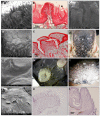
Images from Lemur catta (superior row), Varecia variegata (middle row) and Eulemur macaco (inferior row) showing the dorsum of the tongue (A,E,I) and the zones of the following papillae: circumvallate together with conical (B,F,J), foliate (C,G,K), and fungiform together with filiform (D,H,L). In the left column, the minor division of the scale corresponds to one millimeter.
Figure 2
Figure 2
Ventral aspect of tongue and sublingua in Lemur catta (AC), Varecia variegata (DF) and Eulemur macaco (GI). The sublingua tip (B,C,E,F,H,I) was magnified in several specimens per species. The sublingual tips in the flower-feeding species are wider and more feathered in appearance compared with Lemur catta. The minor division of the scale corresponds to one millimeter.
Figure 3
Figure 3
Images from Lemur catta (AC), Varecia variegata (DF) and Eulemur macaco (GI) tongue’s tip. In all images the ventral surface is observed in the inferior part of the picture. The tip in the flower-feeding species present bigger and modified papillae (Varecia variegata) or is like a brush with long filiform-like papillae. In Lemur catta there are no differences in papillae morphology or distribution when compared with the rest of the tongue’s edge. In image (D), the minor division of the scale corresponds to one millimeter.
Figure 4
Figure 4
Photocomposition of a frontal section of the tongue’s body at the level of the anterior third (images A,D,G) or posterior third (images C,F,I) in Lemur catta (images AC), Varecia variegata (images DF) and Eulemur macaco (images GI). Images (B,D,H) shown a magnification of the area framed in images (A,D,G), focusing on the encapsulated organ found in the antero-ventral position of the tongue in all species studied. Arrow in image (B) points to the presence of an cartilage only in Lemur catta. All sections were stained with hematoxylin—eosin. Scale bar 1 mm.
Figure 5
Figure 5
Images that illustrate the encapsulated structure present in the antero-ventral part of the tongue. Images (AC,E,I) correspond to Lemur catta. Images (D,F,H) correspond to Varecia variegata. Image (G) correspond to Eulemur macaco. Images (AD) (Masson trichrome stain) are from sagital-oriented longitudinal sections. Images (E) (S100 immunostain) and (FI) (hematoxylin—eosin stain) are from frontal-oriented transversal sections. Some images, for descriptive purposes, have the following abbreviations: INF (inferior); SUP (superior); NF (nerve fiber); Ad (adipocyte); Cg (collagen); MF (muscle fiber); Ve (vascular element); CT (cartilaginous tissue) and Cp (capsule). In image (A) part of the encapsulated structure is remarked. Image (C) shows details of the tissue boxed in image (B). Image (D) shows a panoramic of the position of the encapsulated structure (arrow). Images captured with the following objectives: A, 10×; B, 20×; C, 40×; D, 4×; EG, 10×; H,I, 20×.
Figure 6
Figure 6
All images correspond to Lemur catta papillae. SEM images can be observed of fungiform and filiform (A), circumvallate and conical (D), and foliate and conical (G,H). Also presented are Masson trichrome-stained images of filiform (B), conical (C), circumvallate surrounded by conical (E) and a fungiform papilla displaying a taste bud at the top (F). Image (I) clearly shows the Y-shaped organization of circumvallate papillae. A width of one millimeter has the minor division in image (I).
Figure 7
Figure 7
All images correspond to Varecia variegata papillae. SEM images can be observed of fungiform together with filiform (A), foliate (C), circumvallate (D,F), surrounded by flattened conical with several tips and some filiform in the inferior part of the image, a circumvallate central part (G, magnified from image D circumvallate papilla) and a superior view of hammerhead or maze-shaped papillae present on the tip of the tongue (J). Also presented are Masson trichrome-stained images of filiform and fungiform (B) and a circumvallate surrounded by conical (E). Images (IL) illustrate the hammerhead or maze-shaped papillae present on the tip of the tongue, with K and L being sagittal sections stained with hematoxylin—eosin. Image (F) (a superior view of the root of the tongue) shows the Y-shaped disposition of circumvallate papillae and image H displays the crest shape of the flattened conical papillae that surround circumvallate ones in Varecia variegata. A scale of one millimeter in width, can be observed on image (H) (inferior left angle) and in image (F), the minor division is also one millimeter.
Figure 8
Figure 8
All images correspond to Eulemur macaco papillae. SEM images can be observed of fungiform and filiform (A), a conical-like papilla present on the tip of the tongue (C, magnified from Figure 3—image I right inferior angle), circumvallate and conical (F) and foliate (I). Also presented are Masson trichrome-stained images of filiform (B) and circumvallate (E) papillae. Optical images of circumvallate papillae (D, from the posterior branch of the Y-shaped disposition) and foliate papillae (G,H). Image (D,F) correspond to the same sample. The minor division of the scale in image (G) corresponds to one millimeter.

Similar articles

Cited by

References

    1. Jordano P. Coevolution in multispecific interactions among free-living species. Evol. Educ. Outreach. 2010;3:40–46. doi: 10.1007/s12052-009-0197-1. - DOI
    1. Thompson J.N. Four central points about coevolution. Evol. Educ. Outreach. 2010;3:7–13. doi: 10.1007/s12052-009-0200-x. - DOI
    1. Dew J.L., Wright P. Frugivory and seed dispersal by four species of primates in Madagascar’s eastern rain forest. Biotropica. 1998;30:425–437. doi: 10.1111/j.1744-7429.1998.tb00076.x. - DOI
    1. Chapman C.A., Onderdonk D.A. Forests without primates: Primate/plant codependency. Am. J. Primatol. 1998;45:127–141. doi: 10.1002/(SICI)1098-2345(1998)45:1<127::AID-AJP9>3.0.CO;2-Y. - DOI - PubMed
    1. Sussman R.W., Raven P.H. Pollination by lemurs and marsupials: An archaic coevolutionary system. Science. 1978;200:731–736. doi: 10.1126/science.200.4343.731. - DOI - PubMed

LinkOut - more resources