iBet uBet web content aggregator. Adding the entire web to your favor.


Link to original content: https://pubmed.ncbi.nlm.nih.gov/33996277
Cranial anatomy of Besanosaurus leptorhynchus Dal Sasso & Pinna, 1996 (Reptilia: Ichthyosauria) from the Middle Triassic Besano Formation of Monte San Giorgio, Italy/Switzerland: taxonomic and palaeobiological implications - PubMed Skip to main page content
U.S. flag

An official website of the United States government

Dot gov

The .gov means it’s official.
Federal government websites often end in .gov or .mil. Before sharing sensitive information, make sure you’re on a federal government site.

Https

The site is secure.
The https:// ensures that you are connecting to the official website and that any information you provide is encrypted and transmitted securely.

Access keys NCBI Homepage MyNCBI Homepage Main Content Main Navigation
. 2021 May 6:9:e11179.
doi: 10.7717/peerj.11179. eCollection 2021.

Cranial anatomy of Besanosaurus leptorhynchus Dal Sasso & Pinna, 1996 (Reptilia: Ichthyosauria) from the Middle Triassic Besano Formation of Monte San Giorgio, Italy/Switzerland: taxonomic and palaeobiological implications

Affiliations

Cranial anatomy of Besanosaurus leptorhynchus Dal Sasso & Pinna, 1996 (Reptilia: Ichthyosauria) from the Middle Triassic Besano Formation of Monte San Giorgio, Italy/Switzerland: taxonomic and palaeobiological implications

Gabriele Bindellini et al. PeerJ. .

Abstract

Besanosaurus leptorhynchus Dal Sasso & Pinna, 1996 was described on the basis of a single fossil excavated near Besano (Italy) nearly three decades ago. Here, we re-examine its cranial osteology and assign five additional specimens to B. leptorhynchus, four of which were so far undescribed. All of the referred specimens were collected from the Middle Triassic outcrops of the Monte San Giorgio area (Italy/Switzerland) and are housed in various museum collections in Europe. The revised diagnosis of the taxon includes the following combination of cranial characters: extreme longirostry; an elongate frontal not participating in the supratemporal fenestra; a prominent 'triangular process' of the quadrate; a caudoventral exposure of the postorbital on the skull roof; a prominent coronoid (preglenoid) process of the surangular; tiny conical teeth with coarsely-striated crown surfaces and deeply-grooved roots; mesial maxillary teeth set in sockets; distal maxillary teeth set in a short groove. All these characters are shared with the holotype of Mikadocephalus gracilirostris Maisch & Matzke, 1997, which we consider as a junior synonym of B. leptorhynchus. An updated phylogenetic analysis, which includes revised scores for B. leptorhynchus and several other shastasaurids, recovers B. leptorhynchus as a basal merriamosaurian, but it is unclear if Shastasauridae form a clade, or represent a paraphyletic group. The inferred body length of the examined specimens ranges from 1 m to about 8 m. The extreme longirostry suggests that B. leptorhynchus primarily fed on small and elusive prey, feeding lower in the food web than an apex predator: a novel ecological specialisation never reported before the Anisian in a large diapsid. This specialization might have triggered an increase of body size and helped to maintain low competition among the diverse ichthyosaur fauna of the Besano Formation.

Keywords: Besano Formation; Cranial anatomy; Ichthyosauria; Longirostry; Marine reptiles; Middle Triassic; Monte San Giorgio; Osteology; Phylogeny; Shastasauridae.


Conflict of interest statement

Cristiano Dal Sasso is an employee of the Museo di Storia Naturale di Milano, Italy. Torsten Michael Scheyer is an employee of the Paläontologisches Institut und Museum, Universität Zürich, Switzerland.

Figures

Figure 1
Figure 1. Relevant fossil sites in the Monte San Giorgio area.
Map of the Monte San Giorgio area showing the Middle Triassic carbonate succession, the major paleontological quarries in the area (white circles), and the sites of origin of the specimens described in this paper (yellow rhombuses).
Figure 2
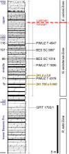
Figure 2. Stratigraphic log of the Besano Formation.
Stratigraphic log of the Besano Formation at the Mirigioli/Punkt 902 outcrop, with the known stratigraphic positions of the specimens described. The stratigraphic position of PIMUZ T 1895 and GPIT 1793/1 is more uncertain, thus expressed by a range line. Log modified from Brack et al. (2005); dating (in millions of years) of layer 71 from Mundil et al. (1996); dating of Tc Tuffs (layers 66–68) from Wotzlaw, Brack & Storck (2017).
Figure 3
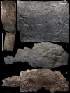
Figure 3. The most complete skeletons of Besanosaurus leptorhynchus.
The most complete skeletons referable to Besanosaurus leptorhynchus. (A) PIMUZ T 1895; (B) BES SC 999; (C) PIMUZ T 4376 (with a Mixosaurus specimen above it); (D) PIMUZ T 4847. Scale bars represent 50 cm.
Figure 4
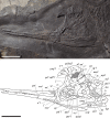
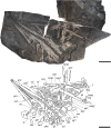
Figure 4. Skull and mandible of Besanosaurus leptorhynchus holotype.
Skull and mandible of Besanosaurus leptorhynchus holotype BES SC 999, and their interpretative drawings. Grey dashed lines and grey labels indicate elements not visible on the surface, grey areas indicate background sediment, light grey areas indicate background bone. Abbreviations: see text. Scale bar represents 10 cm.
Figure 5
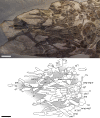
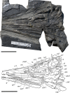
Figure 5. Skull and mandible of PIMUZ T 4376.
Skull and mandible of PIMUZ T 4376, and their interpretative drawings. Grey dashed lines and grey labels indicate elements not visible on the surface, grey areas indicate background sediment, light grey areas indicate background bone. Abbreviations: see text. Scale bar represents 10 cm.
Figure 6
Figure 6. Skull and mandible of PIMUZ T 1895.
Skull and mandible of PIMUZ T 1895, and their interpretative drawings. Grey dashed lines indicate portions of missing elements preserved as counterprints, grey areas indicate background sediment, light grey areas indicate background bone. Abbreviations: see text. Scale bar represents 10 cm.
Figure 7
Figure 7. Skull and mandible of PIMUZ T 4847.
Skull and mandible of PIMUZ T 4847, and their interpretative drawings. Grey areas indicate background sediment. Abbreviations: see text. Scale bar represents 10 cm.
Figure 8
Figure 8. Skull and mandible of GPIT 1793/1.
Skull and mandible of GPIT 1793/1, and their interpretative drawings. Grey dashed lines and grey labels indicate elements not visible on the surface, grey areas indicate background sediment, light grey areas indicate background bone. Abbreviations: see text. Scale bar represents 10 cm.
Figure 9
Figure 9. Skull and mandible of BES SC 1016.
Skull and mandible of BES SC 1016, and their interpretative drawings. Grey areas indicate background sediment, light grey areas indicate background bone. Abbreviations: see text. Scale bar represents 10 cm.
Figure 10
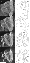
Figure 10. CT slices of BES SC 999 postnarial region.
Selected (most informative) CT slices of the postnarial region of the holotype of Besanosaurus leptorhynchus (BES SC 999), ordered by depth (top to bottom, from the deepest to the most surface level). (A) original slices; (B) slices interpretations. Abbreviations: see text. Scale bar represents 10 cm.
Figure 11
Figure 11. External naris of Besanosaurus leptorhynchus.
External naris and perinarial region of Besanosaurus leptorhynchus. (A) PIMUZ T 1895; (B) BES SC 1016. The white arrows point to the rostral tip of the external naris in both specimens. Scale bars represent 5 cm .
Figure 12
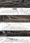
Figure 12. Maxillae of Besanosaurus leptorhynchus.
Maxillae referable to Besanosaurus leptorhynchus. (A and A′) BES SC 999; (B and B′) GPIT 1793/1; (C and C′) PIMUZ T 4376. Light gray areas highlight the maxilla, darker gray areas pinpoint the external naris, when preserved. Abbreviations: see text. Scale bars represent 2.5 cm.
Figure 13
Figure 13. GPIT 1793/1 under UV light.
GPIT 1793/1, partly under visible, partly under UV light (365 nm), highlighting the sutures, otherwise not easily distinguishable from fractures, of frontals and adjacent bones on the ventral (internal) side of the skull roof: the extent of the frontals is greater internally than externally (see Figs. 4–6). Scale bar represents 5 cm.
Figure 14
Figure 14. Braincase elements of Besanosaurus leptorhynchus.
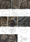
Disarticulated braincase elements of Besanosaurus leptorhynchus GPIT 1793/1 (A–C, G), PIMUZ T 4376 (D and E), and BES SC 999 (F). Interpretative drawings are denoted by apostrophes. (A and A′) supraoccipital in posterior view; (B and B′) opisthotic in rostromedial view. (C and C′) prootic in caudal view. (D and D′) exoccipital in rostral? view; (E and E′) basioccipital in dorsal view; (F and F′) basioccipital in caudal view; (G and G′) parabasisphenoid in ventral view. Dashed lines indicate portions of bones not visible on the surface, thin lines indicate bone structures. Abbreviations: see text. Scale bars represent 1 cm.
Figure 15
Figure 15. CT image of BES SC 1016.
CT image of specimen BES SC 1016. Note the pterygoids still in articulation and the relatively narrow interpterygoid vacuity (black arrow). The caudal extremities of the pterygoids are marked by grey arrows (full extent of the pterygoids is visible in Fig. 9). Note also the implantation of the mesialmost (caudalmost) premaxillary teeth, that are nested in separate sockets (white arrow). Scale bar represents 10 cm.
Figure 16
Figure 16. Jugal and quadratojugal of GPIT 1793/1.
Specimen GPIT 1793/1, left jugal in medial view (top) and right quadratojugal in lateral view (bottom). Dashed lines indicate portions of missing elements preserved as counterprints, thin lines indicate bone depressions. Abbreviations: see text. Scale bar represents 5 cm.
Figure 17
Figure 17. Quadrates of Besanosaurus leptorhynchus.
Quadrates of Besanosaurus leptorhynchus. (A) right quadrate of PIMUZ T 4376 in rostrolateral (anterior) view; (B) CT scan of the left quadrate area of BES SC 999; (C) left quadrate of BES SC 999 in rostrolateral view, under visible light; (D) right quadrate of GPIT 1793/1 in rostrolateral view; (E) right quadrate of PIMUZ T 4847 in rostrolateral view; the upper half of the quadrate is missing, as well as most of its lateral portion. Interpretative drawings below. Dashed lines indicate portions of missing elements, thin lines indicate bone structures. Abbreviations: see text. Scale bar represents 5 cm.
Figure 18
Figure 18. Teeth of Besanosaurus leptorhynchus.
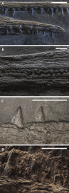
Teeth of the specimens referable to Besanosaurus leptorhynchus. (A) PIMUZ T 4376: rostralmost (mesialmost) teeth of the dentary (above, turned upside down) and the premaxillae (below); (B) BES SC 999: premaxillary teeth at mid-length of the rostrum; (C) GPIT 1793/1: rostralmost teeth of the right maxilla; (D) PIMUZ T 4847: ?dentary teeth at mid-length of the rostrum. Scale bars represent 1 cm.
Figure 19
Figure 19. Selected plots showing relevant cranial ratios across studied specimens.
Selected plots showing relevant cranial ratios across studied specimens supporting that they represent an ontogenetic series. (A) Presacral length/jaw length; (B) orbital rostrocaudal diameter/Jaw length; (C) jaw length/orbital rostrocaudal diameter.
Figure 20
Figure 20. Cranial reconstruction of Besanosaurus leptorhynchus.
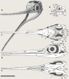
Cranial reconstruction of Besanosaurus leptorhynchus. Articulated skull and mandible in (A) left rostrolateral, (B) caudal (occipital), (C) dorsal, (D) left lateral, and (E) ventral (palatal) view. Abbreviations: see text. Line drawings by Marco Auditore.
Figure 21
Figure 21. Cladogram of Ichthyosauriformes and phylogenetic position of Besanosaurus.
(A) 50% Majority rule consensus of 14,480 MPTs of 713 steps (CI = 0.363, RI = 0.788) obtained from parsimony analysis of the character-taxon matrix of Huang et al. (2019). Note that ‘shastasaurids’ are recovered as a grade at the base of Merriamosauria. Numbers above nodes indicate proportion of MPTs with specific node resolution. (B) Alternative resolution of Merriamosauria, with Shastasauridae recovered as a clade, and Besanosaurus leptorhynchus being the sister taxon of Guizhouichthyosaurus. (C) Alternative resolution of Merriamosauria, with Shastasauridae recovered as a clade, and Besanosaurus leptorhynchus being the earliest-diverging shastasaurid. Abbreviations: Call., Callawayia; Ch., Chaohusaurus; Cymb., Cymbospondylus; Lepto., Leptonectes; Mixo., Mixosaurus; Oph., Ophthalmosaurus; Qian., Qianichthyosaurus; Phal., Phalarodon; Shon., Shonisaurus.
Figure 22
Figure 22. Hooklet of a coleoid cephalopod.
Isolated hooklet of a coleoid cephalopod preserved in the thoracic region of BES SC 999, holotype of Besanosaurus leptorhynchus. Scale bar represents 1 mm.
Figure 23
Figure 23. Early and Middle Triassic ichthyopterygian heads possessing longirostry.
Simplified outlines of four different Early and Middle Triassic ichthyopterygian heads possessing a long and slender rostrum. Specimens are at the same scale. (A) Utatsusaurus hataii (UHR 30691, Motani, Minoura & Ando, 1998); (B) Grippia longirostris (PMU R445, Motani, 2000); (C) Mixosaurus cornalianus (BES SC 1000, Renesto et al., 2020); (D) Besanosaurus leptorhynchus (PIMUZ T 4847, this paper); (E) Cymbospondylus buchseri (PIMUZ T 4351, Sander, 1989). Scale bar represents 10 cm.

Similar articles

Cited by

References

    1. Andrews CW. A descriptive catalogue of the marine reptiles of the Oxford Clay, part 1. London: British Natural History Museum; 1910. p. 215.
    1. Benton MJ, Zhang Q, Hu S, Chen ZQ, Wen W, Liu J, Huang J, Zhou C, Xie T, Tong J, Choo B. Exceptional vertebrate biotas from the Triassic of China, and the expansion of marine ecosystems after the Permo-Triassic mass extinction. Earth Sciences Review. 2013;125(5):199–243. doi: 10.1016/j.earscirev.2013.05.014. - DOI
    1. Bernasconi SM. Geochemical and microbial controls on dolomite formation and organic matter production/preservation in anoxic environments a case study from the Middle Triassic Grenzbitumenzone, Southern Alps (Ticino, Switzerland) 1991. p. 196. D. Phil. thesis, Swiss Federal Institute Of Technology Zürich, Switzerland,
    1. Bernasconi SM. Geochemical and microbial controls on dolomite formation in anoxic environments: a case study from the Middle Triassic (Ticino, Switzerland) Contributions to Sedimentology. 1994;19:1–109.
    1. Bernasconi SM, Riva A. Organic geochemistry and depositional environment of a hydrocarbon source rock: the Middle Triassic Grenzbitumenzone Formation, Southern Alps, Italy/Switzerland. In: Spencer AM, editor. Generation, Accumulation and Production of Europe’s Hydrocarbons. Vol. 3. Berlin: Springer; 1993. pp. 179–190.

Grants and funding

Comparative data collection by Andrzej S. Wolniewicz was funded by a Natural Environment Research Council (NERC) Ph. D. Studentship (cohort grant NE/L501530/1) carried out at the Department of Earth Sciences, University of Oxford (2013–2017). Torsten M. Scheyer was supproted by the Swiss National Science Foundation (grant no. 31003A_179401). The funders had no role in study design, data collection and analysis, decision to publish, or preparation of the manuscript.

LinkOut - more resources