iBet uBet web content aggregator. Adding the entire web to your favor.


Link to original content: https://pubmed.ncbi.nlm.nih.gov/33395550
Biophysics of Chromatin Remodeling - PubMed Skip to main page content
U.S. flag

An official website of the United States government

Dot gov

The .gov means it’s official.
Federal government websites often end in .gov or .mil. Before sharing sensitive information, make sure you’re on a federal government site.

Https

The site is secure.
The https:// ensures that you are connecting to the official website and that any information you provide is encrypted and transmitted securely.

Access keys NCBI Homepage MyNCBI Homepage Main Content Main Navigation
Review
. 2021 May 6:50:73-93.
doi: 10.1146/annurev-biophys-082520-080201. Epub 2021 Jan 4.

Biophysics of Chromatin Remodeling

Affiliations
Review

Biophysics of Chromatin Remodeling

Ilana M Nodelman et al. Annu Rev Biophys. .

Abstract

As primary carriers of epigenetic information and gatekeepers of genomic DNA, nucleosomes are essential for proper growth and development of all eukaryotic cells. Although they are intrinsically dynamic, nucleosomes are actively reorganized by ATP-dependent chromatin remodelers. Chromatin remodelers contain helicase-like ATPase motor domains that can translocate along DNA, and a long-standing question in the field is how this activity is used to reposition or slide nucleosomes. In addition to ratcheting along DNA like their helicase ancestors, remodeler ATPases appear to dictate specific alternating geometries of the DNA duplex, providing an unexpected means for moving DNA past the histone core. Emerging evidence supports twist-based mechanisms for ATP-driven repositioning of nucleosomes along DNA. In this review, we discuss core experimental findings and ideas that have shaped the view of how nucleosome sliding may be achieved.

Keywords: SF2 superfamily; Snf2-type ATPase motor; bulge/loop propagation; histone; nucleosome; twist defect.


Figures

Figure 1.
Figure 1.. The structure of the nucleosome.
(a) Overview of a high-resolution nucleosome crystal structure (PDB code 1KX5; Reference 29). The 147 bp DNA duplex wraps around the histone octamer approximately 1.65 times. The dyad indicates the central two-fold axis of symmetry. (b) With each turn of the DNA duplex, the histone core makes regular interactions with DNA where the phosphate backbone faces the core. Yellow surfaces represent regions of the histone core that make direct hydrogen bonding and van der Waals contacts with the DNA backbone. (c) DNA sites around the nucleosome are referred to by their superhelix location (SHL). SHL0 defines where the inward-facing major groove aligns with the central two-fold or dyad axis of the nucleosome, and each successive inward-facing major or minor groove is given integer (major) or half integer (minor) values (80).
Figure 2.
Figure 2.. Twist defects on the nucleosome.
(a) The twist diffusion model for nucleosome repositioning (123, 124). A corkscrew shift of the duplex causes a DNA segment between two minor groove contacts to absorb an additional bp, which locally changes DNA twist (a twist defect). Once formed, a corkscrew shift on the other side transfers the twist defect further onto the nucleosome, accompanied by a 1 bp translation of DNA. (b) Changes in DNA geometry accompanying twist defects. Comparison of the two sides of the original 146 bp nucleosome structure at SHL2 shows that the one bp difference is distributed ~5 nt away on the two strands, farthest from histone-DNA contacts. (c) As highlighted by rectangular boxes, the shift in the DNA backbone at the farthest point from the histone core correlates with bp tilting. (d) Two nucleosome crystal structures that captured twist defects at two separated locations, SHL2 and SHL5 (80, 117). These nucleosomes had the same DNA sequence and length, only differing by the presence or absence of a pyrrole-imidazole polyamide at SHL4 (1M18 structure). Red arrows represent expected corkscrew rotations of intervening DNA needed for a twist defect to collapse at one location and appear at the other.
Figure 3.
Figure 3.. Bulge/loop propagation models of nucleosome sliding.
(a) A loop recapture model. In this model, DNA unwraps and then rewraps with a shifted position, trapping a loop of DNA. Adapted from Längst & Becker 2004 (67). (b) Nucleosome repositioning resulting from transcription by an RNA polymerase around the histone core. Adapted with permission from Studitsky et al., 1997 (114). (c) A model for how the SWI/SNF remodeler alters nucleosome structure, creating persistent DNA loops on the histone surface. Adapted from Fan et al., 2003 (37). (d) A loop propagation model where the entry-side loop is created by cooperation between the ATPase motor at SHL2 (Tr) and a DNA-binding domain (DBD) at the nucleosome edge. DNA movement past the ATPase motor occurs between states 2 and 3, highlighted by the shift of an asterisk (*) reference point. Adapted from Clapier & Cairns, 2009 (25). (e) A model where translocation of the RSC ATPase motor (Tr) at the internal SHL2 site directly spools out DNA into a loop at the dyad. Adapted from Zhang et al., 2006 (141).
Figure 4.
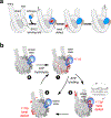
Figure 4.. Models of nucleosome sliding via twist defects.
(a) Overview of remodeler-catalyzed DNA motions, showing that DNA first shifts onto the nucleosome from the entry side, is transiently absorbed on the nucleosome after being expelled from SHL2, and then shifts off the exit side. (b) Possible pathways for how remodeler ATPases stimulate 1 bp shifts of DNA around the nucleosome.
Figure 5.
Figure 5.. Contacts and structural changes associated with the open state ATPase at SHL2.
(a) In the open state, the shifted position of ATPase lobe 2 on the nucleosome alters the path for the tracking strand, which correlates with a change in register (red ovals) of the tracking strand (orange) but not guide (yellow). (b) Due to the shift in register of the tracking strand, DNA base pairs show a marked tilting, which continues outside the SHL2 binding site. (c) Base pair tilting originates in the ATPase motor. Superpositioning lobe 2 from a closed ATPase (gray, 6JYL) onto that of an open ATPase (colored, 6IRO) shows a similar path of the DNA backbone even though the register of the two strands differs by 1 nt, evident as bp tilting. This is consistent with a change in DNA geometry due to contacts made by both ATPase lobes. (d) Snf2-specific insertions on lobe 2 contact the histone core. In most nucleosome-bound structures, the polypeptide segment immediately preceding the gating helix contacts residues on the L1 loop of histone H3 (sticks). The loop prior to this segment is often disordered, however in nucleosome-bound complexes of Chd1 (116), CHD4 (38), and SNF2h (6), this loop was ordered and appeared to make more extensive contacts with histone H3.
Figure 6.
Figure 6.. SWR1 distorts DNA on the nucleosome.
(a) Overview of the SWR1-nucleosome complex, bound with ADP·BeF3 (132). (b) Comparison of DNA conformations at SHL2. Histone and remodeler proteins are omitted for clarity. (c) A kink in the guide strand alters the path of the DNA duplex in the SWR1 complex. Shown is a surface/backbone representation of the SWR1 ATPase motor (tan, blue; 6GEJ) bound to a segment of DNA surrounding SHL2. The position of the gray DNA results from superimposing the ATPase motor of an ISWI remodeler (6JYL), also bound to ADP·BeF3, with the SWR1 ATPase motor (ISWI ATPase motor omitted for clarity). (d) Comparison of the gating helices of Snf2-insertions for remodelers bound to nucleosomes (solid) to the same regions of the nucleosome-free structures (red outlines). For each structure, superpositioning ATPase lobe 2 from unbound structures onto the nucleosome complex produces a steric clash with the guide strand. (e) The SWR1 ATPase lifts the DNA backbone away from SHL2.5, partially disrupting histone contacts. This shifts the minor groove away from H3 arginine 83, which appears to no longer be inserted (not shown). The nucleosome alone structure (white, 1KX5) is shown for comparison.

Similar articles

Cited by

References

    1. Aalfs JD, Narlikar GJ, Kingston RE. 2001. Functional differences between the human ATP-dependent nucleosome remodeling proteins BRG1 and SNF2H. J. Biol. Chem. 276:34270–8 - PubMed
    1. Alfert A, Moreno N, Kerl K. 2019. The BAF complex in development and disease. Epigenetics Chromatin. 12:19,019–0264-y - PMC - PubMed
    1. Allis CD, Jenuwein T. 2016. The molecular hallmarks of epigenetic control. Nat. Rev. Genet. 17:487–500 - PubMed
    1. Aoyagi S, Hayes JJ. 2002. hSWI/SNF-catalyzed nucleosome sliding does not occur solely via a twist-diffusion mechanism. Mol. Cell. Biol. 22:7484–90 - PMC - PubMed
    1. Aoyagi S, Wade PA, Hayes JJ. 2003. Nucleosome sliding induced by the xMi-2 complex does not occur exclusively via a simple twist-diffusion mechanism. J. Biol. Chem. 278:30562–8 - PubMed

Publication types

MeSH terms

LinkOut - more resources