iBet uBet web content aggregator. Adding the entire web to your favor.


Link to original content: https://pubmed.ncbi.nlm.nih.gov/33203861
Structure of the human sodium leak channel NALCN in complex with FAM155A - PubMed Skip to main page content
U.S. flag

An official website of the United States government

Dot gov

The .gov means it’s official.
Federal government websites often end in .gov or .mil. Before sharing sensitive information, make sure you’re on a federal government site.

Https

The site is secure.
The https:// ensures that you are connecting to the official website and that any information you provide is encrypted and transmitted securely.

Access keys NCBI Homepage MyNCBI Homepage Main Content Main Navigation
. 2020 Nov 17;11(1):5831.
doi: 10.1038/s41467-020-19667-z.

Structure of the human sodium leak channel NALCN in complex with FAM155A

Affiliations

Structure of the human sodium leak channel NALCN in complex with FAM155A

Jiongfang Xie et al. Nat Commun. .

Abstract

NALCN, a sodium leak channel expressed mainly in the central nervous system, is responsible for the resting Na+ permeability that controls neuronal excitability. Dysfunctions of the NALCN channelosome, NALCN with several auxiliary subunits, are associated with a variety of human diseases. Here, we report the cryo-EM structure of human NALCN in complex with FAM155A at an overall resolution of 3.1 angstroms. FAM155A forms extensive interactions with the extracellular loops of NALCN that may help stabilize NALCN in the membrane. A Na+ ion-binding site, reminiscent of a Ca2+ binding site in Cav channels, is identified in the unique EEKE selectivity filter. Despite its 'leaky' nature, the channel is closed and the intracellular gate is sealed by S6I, II-III linker and III-IV linker. Our study establishes the molecular basis of Na+ permeation and voltage sensitivity, and provides important clues to the mechanistic understanding of NALCN regulation and NALCN channelosome-related diseases.


Conflict of interest statement

The authors declare no competing interests.

Figures
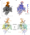
Fig. 1
Fig. 1. Cryo-EM structures of human NALCN in complex with FAM155A.
a Cryo-EM map of human NALCN in complex with FAM155A. NALCN and FAM155A are colored gray and orange, respectively. The map was contoured at 0.8 threshold level in ChimeraX. b Local resolution map estimated by cryoSPARC and generated in Chimera. c Overall structure of the NALCN-FAM155A complex presented in two side views. The protein structure is shown in cartoon. NALCN is domain colored with repeats I, II, III, IV, and the CTD in light gray, green, yellow, cyan, and slate, respectively. The III-IV linker and FAM155A are colored orange. Lipid molecules and the sugar moieties at the glycosylation sites are shown as sticks. The color schemes are applied in all figures. All structure figures were prepared with PyMOL.
Fig. 2
Fig. 2. Structural comparisons between NALCN and Nav/Cav channels.
a Overall structures of human NALCN, human Nav1.7 (PDB: 6J8I), and rabbit Cav1.1 (PDB: 6JP5). The ion conducting subunits of Nav1.7 and Cav1.1 are colored slate and light pink, respectively. The auxiliary subunits β1 and β2 of Nav1.7 are colored cyan and magenta, and the auxiliary subunits α2δ, β, γ of Cav1.1 are colored cyan, yellow, and green, respectively. b FAM155A adopts a unique binding site on NALCN that differs from the auxiliary subunits of Nav/Cav channels. The structure of NALCN is superimposed with Nav1.7 or Cav1.1 based on the ion-conducting subunit of each complex. Nav1.7 is shown in an extracellular view (left), and Cav1.1 is shown in a side view (right). c Structural features in repeat I and a unique lipid-binding site of NALCN. The S4-5I linker of NALCN is a loop instead of a helix in Nav1.7/Cav1.1. S5I in NALCN is extended three-helix turns into the cytosol compared to that of Nav1.7/Cav1.1. An identified lipid molecule in NALCN occupies a position that corresponds to the S4-5 helix in Nav1.7/Cav1.1. Local structural deviations around the lipid between NALCN and Nav1.7/Cav1.1 are indicated by black arrows.
Fig. 3
Fig. 3. Specific interactions between NALCN and FAM155A.
a One-dimensional (1D) schematic view of FAM155A. The region that was resolved in the current structure is colored orange. The position of the glycosylation site and predicted two transmembrane helices, polyQ, and polyG motifs, are labeled. b 2D topological structure of FAM155A. c Interaction interface between NALCN and FAM155A. NALCN is shown in surface and FAM155A is shown in cartoons. Details in the rectangle boxes are presented in d. d Zoom in views of each interaction interface. The electrostatic interactions are indicated by red dashed lines. The residues from NALCN and FAM155A are labeled black and orange, respectively. Please also refer to Supplementary Table 2 for a summary of the interactions between NALCN and FAM155A.
Fig. 4
Fig. 4. Structural features of the VSDs of NALCN.
a Structure-based sequence alignment of the four VSDs. The boundaries for the S1 to S4 segments are shaded gray. The gating charges (GCs) in the S4 helices are colored red. The residues that correspond to the charge transfer center (CTC) on S2 and S3 helices as well as the polar or acidic residues on S1 that coordinate GCs are highlighted in blue. b All four VSDs adopt up or depolarized state. The four VSDs are superimposed relative to CTC and An1 on S2. For visual clarity, the S1 segments are omitted. c Structural details of each VSD. The GCs, CTCs, and the polar or acidic residues participating in GC coordination are shown in sticks. The GCs are labeled in red. The S4-5I loop and non-conserved aromatic residues in the CTC of repeat III are highlighted by red circles. The short 310 helix turn in repeat IV is indicated by a dark green line.
Fig. 5
Fig. 5. The EEKE selectivity filter of NALCN.
a Overall structure of the pore domain of NALCN. Side views of the pore domain from the diagonal repeats are shown. The EEKE residues in the selectivity filter (SF) are shown in stick. Repeat I and repeat III adopt large extracellular loops, reminiscent of Cav1.1. A π helix appears in each S6 segment and is indicated by red arrows. b Sequence alignment of the SF and the connecting pore helices among NALCN, Cav1.1, and Nav1.4. The critical EEKE (E280, E554, K1115, and E1389) residues in NALCN and the corresponding residues in Cav1.1 (EEEE) and Nav1.4 (DEKA) are shaded yellow. The invariant Trp residues and the highly conserved Thr residues are colored light green. The acidic residues that may help to coordinate cations in NALCN are shaded orange. c Structure comparisons of the SF between NALCN and Nav1.4 (left) or Cav1.1 (right). The critical residues in the SF are shown in the stick. d Top view and two side views of sodium probability density generated from pore domain equilibration. Three potential Na+ binding sites were identified and labeled in purple. The most stable binding site (Site1) is signposted by a Na+ ion (magenta sphere). Please also refer to Supplementary Movie for the illustrative trajectory of equilibration. e Ion numbers within 4 Å of SF residues (EEKE) over time (left) and the probability statistics (right). The statistics represent the results of three independent simulations. f Structural comparison of Site 3 (EED motif) in NALCN and the Na+ binding site (DEE motif) in Nav1.4 (top) and of Site1 in NALCN and a Ca2+ binding site in Cav1.1 (bottom). Na+ and Ca2+ are shown as purple and green spheres, respectively.
Fig. 6
Fig. 6. A closed intracellular pore.
a Ion permeation path of NALCN. The pore region of NALCN is shown in cartoon and FAM155A is shown in the surface. The ion permeation path calculated by HOLE is illustrated by purple dots on the left. The extracellular ion entrance is indicated by a red arrow. The details in the rectangular region are shown in b. The corresponding pore radii along the permeation path of human NALCN (black), Nav1.4 (slate), and Cav1.1(pink) are compared in the middle. Two narrowest layers of the intracellular gate are presented in extracellular views on the right. b Zoomed views of the intracellular gate. The electrostatic interactions are indicated by red dashed lines. The residues from S6I, II-III linker, and III-IV linker are colored black, green, and orange, respectively. c Pore structure comparisons between NALCN and Nav1.4 (top) or Cav1.1 (bottom). NALCN is domain colored, and Nav1.4 and Cav1.1 are colored in slate and light pink, respectively. An intracellular view and a side view are shown for each comparison. The major conformational changes from Nav1.4 or Cav1.1 to NALCN are indicated by red arrows.
Fig. 7
Fig. 7. Disease mutations mapping on NALCN and TTX incompatibility of NALCN.
a Please also refer to Supplementary Table 3 for a summary of the disease-related mutations on human NALCN. The Cα atoms of the residues whose mutations lead to human diseases are mapped to the structure of NALCN and shown as spheres. The mapping is shown on both sides and extracellular views. b Superimposition of the pore helices and SF of NALCN with that of TTX-bound Nav1.7 (left). The determinant Tyr362 for TTX coordination in Nav1.7 is not conserved in NALCN and the structural deviations in the pore region of NALCN also result in potential clashes with the TTX binding site, making NALCN and TTX incompatible. As a comparison, despite lack of the determinant Tyr, TTX-insensitive Nav1.5 shows a similar backbone structure of pore helices and SF to that of TTX-bound Nav1.7, thus retains a lower binding affinity to TTX (right).

Similar articles

Cited by

References

    1. Bean BP. The action potential in mammalian central neurons. Nat. Rev. Neurosci. 2007;8:451–465. doi: 10.1038/nrn2148. - DOI - PubMed
    1. Ren DJ. Sodium leak channels in neuronal excitability and rhythmic behaviors. Neuron. 2011;72:899–911. doi: 10.1016/j.neuron.2011.12.007. - DOI - PMC - PubMed
    1. Catterall WA. The molecular basis of neuronal excitability. Science. 1984;223:653–661. doi: 10.1126/science.6320365. - DOI - PubMed
    1. Lu B, et al. The neuronal channel NALCN contributes resting sodium permeability and is required for normal respiratory rhythm. Cell. 2007;129:371–383. doi: 10.1016/j.cell.2007.02.041. - DOI - PubMed
    1. Lee JH, Cribbs LL, Perez-Reyes E. Cloning of a novel four repeat protein related to voltage-gated sodium and calcium channels. FEBS Lett. 1999;445:231–236. doi: 10.1016/S0014-5793(99)00082-4. - DOI - PubMed

Publication types