iBet uBet web content aggregator. Adding the entire web to your favor.


Link to original content: https://pubmed.ncbi.nlm.nih.gov/30772942/
Cranial anatomy of the early cynodont Galesaurus planiceps and the origin of mammalian endocranial characters - PubMed Skip to main page content
U.S. flag

An official website of the United States government

Dot gov

The .gov means it’s official.
Federal government websites often end in .gov or .mil. Before sharing sensitive information, make sure you’re on a federal government site.

Https

The site is secure.
The https:// ensures that you are connecting to the official website and that any information you provide is encrypted and transmitted securely.

Access keys NCBI Homepage MyNCBI Homepage Main Content Main Navigation
. 2019 May;234(5):592-621.
doi: 10.1111/joa.12958. Epub 2019 Feb 17.

Cranial anatomy of the early cynodont Galesaurus planiceps and the origin of mammalian endocranial characters

Affiliations

Cranial anatomy of the early cynodont Galesaurus planiceps and the origin of mammalian endocranial characters

Luisa C Pusch et al. J Anat. 2019 May.

Abstract

The cranial anatomy of the early non-mammalian cynodont Galesaurus planiceps from the South African Karoo Basin is redescribed on the basis of a computed tomographic reconstruction of the skull. Previously, little was known about internal skull morphology and the nervous and sensory system of this taxon. The endocranial anatomy of various cynodonts has been intensively studied in recent years to understand the origin of mammalian characters in the nasal capsule, brain and ear. However, these studies have focused on only a few taxa, the earliest of which is another Early Triassic cynodont, Thrinaxodon liorhinus. Galesaurus is phylogenetically stemward of Thrinaxodon and thus provides a useful test of whether the mammal-like features observed in Thrinaxodon were present even more basally in cynodont evolution. The cranial anatomy of G. planiceps is characterized by an intriguing mosaic of primitive and derived features within cynodonts. In contrast to the very similar internal nasal and braincase morphology of Galesaurus and Thrinaxodon, parts of the skull that seem to be fairly conservative in non-prozostrodont cynodonts, the morphology of the maxillary canal differs markedly between these taxa. Unusually, the maxillary canal of Galesaurus has relatively few ramifications, more similar to those of probainognathian cynodonts than that of Thrinaxodon. However, its caudal section is very short, a primitive feature shared with gorgonopsians and therocephalians. The otic labyrinth of Galesaurus is generally similar to that of Thrinaxodon, but differs in some notable features (e.g. proportional size of the anterior semicircular canal). An extremely large, protruding paraflocculus of the brain and a distinct medioventrally located notch on the anterior surface of the tabular, which forms the dorsal border of the large parafloccular lobe, are unique to Galesaurus among therapsids with reconstructed endocasts. These features may represent autapomorphies of Galesaurus, but additional sampling is needed at the base of Cynodontia to test this.

Keywords: bony labyrinth; brain endocast; endocranial anatomy; mammals; micro-computed tomography; synapsids.


Figures

Figure 1
Figure 1
Phylogeny of Cynodontia showing important taxa discussed in this paper. Modified from Abdala (2007) and Ruta et al. (2013).
Figure 2
Figure 2
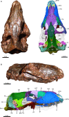
Skull of AMNH FARB 2227. (A) Photograph (left) and 3D reconstruction (right) of the skull in dorsal view. (B) Photograph (top) and 3D reconstruction (bottom) of the skull in right lateral view. The lower jaw was digitally removed in the reconstructed views. bo, basioccipital; C, upper canine; d, dentary; epi, epipterygoid; fr, frontal; hy, hyoid; ju, jugal; la, lacrimal; mx, maxilla; na, nasal; op, opisthotic; pa, parietal; pbs, parabasisphenoid; pl, palatine; pmx, premaxilla; po, postorbital; pp, postparietal; prf, prefrontal; pro, prootic; pt, pterygoid; q, quadrate; smx, septomaxilla; so, supraoccipital; sq, squamosal; ta, tabular.
Figure 3
Figure 3
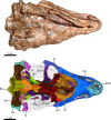
Skull of AMNH FARB 2227 in ventral view. Photograph (top) and 3D reconstruction (bottom). The lower jaw was digitally removed in the reconstructed view. ang, angular; bo, basioccipital; d, dentary; ect, ectopterygoid; epi, epipterygoid; hy, hyoid; ju, jugal; mx, maxilla; op, opisthotic; pbs, parabasisphenoid; pl, palatine; pmx, premaxilla; po, postorbital; pt, pterygoid; q, quadrate; st, stapes; sq, squamosal; vo, vomer.
Figure 4
Figure 4
The rostral nasal cavity of AMNH FARB 2227. (A) 3D reconstruction of the rostral nasal chamber in left anterodorsolateral view. (B,C) Transverse μCT sections through the rostral pars anterior of the nasal cavity. inp, intranarial process; la, lacrimal; mx, maxilla; na, nasal; pdp, posterodorsal process of the septomaxilla; pmx, premaxilla; pnp, prenasal process of the premaxilla; smx, septomaxilla; smxc, septomaxillary canal; vo, vomer.
Figure 5
Figure 5
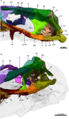
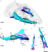
3D reconstructions of the maxillary canal (in green), maxillary antrum (in pink), and the lacrimal (in turquoise) of AMNH FARB 2227: (A) right lateral view with the skull transparent, (B) left anterodorsolateral view with the skull transparent, (C) right posterodorsolateral view with the skull transparent and (D) right lateral view. Dotted line illustrates the caudal extremity of the infraorbital nerve. C, upper canine; cal, caudal alveolar ramus; ena, external nasal rami of the ION; ina; internal nasal ramus of the ION; ION, infraorbital nerve; la, lacrimal; mal, medial alveolar ramus; mxa, maxillary antrum; olac, opening of lacrimal canal; PCs, upper postcanines; ral, rostral alveolar ramus; slr, supralabial rami of the ION.
Figure 6
Figure 6
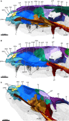
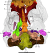
Mid‐sagittal section through the snout of AMNH FARB 2227 to show the internal morphology of the reconstructed nasal region. (A,B) Internal view of the nasal region with the vomer, palatine and frontal digitally removed in (B) to expose the internal structure. (C) Internal morphology of the nasal region in right posterodorsolateral view with the skull roof digitally removed to expose the internal structure. C, upper canine; ect, ectopterygoid; extn, external naris; fr, frontal; inp, intranarial process; ju, jugal; la, lacrimal; mx, maxilla; na, nasal; obl, olfactory bulb location; och, olfactory chamber; olac, opening of lacrimal canal; pa, parietal; PCs, upper postcanines; pcult, processus cultriformis; pl, palatine; pmx, premaxilla; pnp, prenasal process of the premaxilla; po, postorbital; ppmx, palatal process of the maxilla; prf, prefrontal; pt, pterygoid; rch, respiratory chamber; rmxt, ridge for cartilaginous maxilloturbinals; rnt, ridge for cartilaginous nasal turbinals; smx, septomaxilla; vo, vomer.
Figure 7
Figure 7
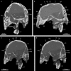
Transverse μCT sections through the respiratory chamber in (A,C,D) and the olfactory chamber (B) of the nasal region of AMNH FARB 2227. d, dentary; ju, jugal; la, lacrimal; lac, lacrimal canal; lr, lateral ridge; mr, median ridge; mx, maxilla; mxa, maxillary antrum; mxc, maxillary canal; na, nasal; np, nasopharyngeal passage; och, olfactory chamber; o‐mxa, opening of maxillary antrum; pl, palatine; ppmx, palatal process of the maxilla; prf, prefrontal; rch, respiratory chamber; tl, transverse lamina; vo, vomer.
Figure 8
Figure 8
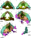
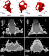
3D reconstruction of the snout of AMNH FARB 2227. (A) Snout in dorsal aspect with the roof, lacrimal and prefrontal digitally removed to expose the internal structure. (B) Ventral view of the palate. C, upper canine; ect, ectopterygoid; I, incisor; if, incisive foramen; ju, jugal; mx, maxilla; PCs, upper postcanines; pl, palatine; pmx, premaxilla; pnp, prenasal process of the premaxilla; ppmx, palatal process of the maxilla; pt, pterygoid; sc, secondary choana; smx, septomaxilla; tl, transverse lamina; VNO, vomeronasal organ; vo, vomer.
Figure 9
Figure 9
3D reconstruction of the skull roof in ventrolateral view. fr, frontal; na, nasal; pa, parietal; po, postorbital; prf, prefrontal; rnt, ridge for cartilaginous nasal turbinals.
Figure 10
Figure 10
Mid‐sagittal section through the reconstructed braincase of AMNH FARB 2227 to show the internal morphology of the sidewalls. (A) Internal view of the sidewalls of the braincase. (B) Internal morphology of the sidewalls of the braincase in right anterodorsolateral view. bo, basioccipital; epi, epipterygoid; fr, frontal; hy, hyoid; jf, jugular foramen; ju, jugal; na, nasal; obl, olfactory bulb location; op, opisthotic; pa, parietal; pan, pila antotica; pbs, parabasisphenoid; pcult, processus cultriformis; pf, parietal foramen; pl, palatine; po, postorbital; pp, postparietal; prf; prefrontal, pro, prootic; pt, pterygoid; q, quadrate; so, supraoccipital; st, stapes; sq, squamosal; ta, tabular; uz, unossified zone; vo, vomer; V, trigeminal nerve; VI, abducens nerve; VII, facial nerve; XII, hypoglossal nerve.
Figure 11
Figure 11
Virtual 3D reconstruction of the floor of braincase with the skull transparent in dorsal view. The right epipterygoid was digitally removed. Asterisk indicates the location of the cavum epiptericum. bo, basioccipital; bpt, basipterygoid process; cf, carotid foramen; cr, cochlear recess; dpq, dorsal plate of the quadrate; ds, dorsum sellae; ect, ectopterygoid; epi, epipterygoid; hy, hyoid; jf, jugular foramen; lfl, lateral flange of the prootic; op, opisthotic; pan, pila antotica; pcult, processus cultriformis; pl, palatine; ppf, pterygoparoccipital foramen; pro, prootic; pt, pterygoid; q, quadrate; qpt; quadrate ramus of the pterygoid; st, stapes; stu, sella turcica; t, trochlea; tf, transverse flange of the pterygoid.
Figure 12
Figure 12
3D reconstructions of the occiput, prootic and middle ear of AMNH FARB 2227. (A) Occiput in anterior view. (B) Posterior view of the occiput. (C) Occiput in anterior aspect with the squamosal and supraoccipital digitally removed to expose the anterior surface of the tabular. (D) Occiput and middle ear in left and right (E) anterodorsolateral aspect. (F) Medial view of the squamosal and the quadrate. ac, anterior crus; bo, basioccipital; dpq, dorsal plate of the quadrate; dps, dorsal process of the stapes; ex, exoccipital; fm, foramen magnum; fp, stapes footplate; hy, hyoid; jf, jugular foramen; ln, lateral notch; npfl, notch for the paraflocculus; op, opisthotic; pan, pila antotica; pc, posterior crus; pp, postparietal; ppf, pterygoparoccipital foramen; pro, prootic; ptf, post‐temporal fenestra; q, quadtrate; so, supraoccipital; sq, squamosal; sqd, squamosal depression; st, stapes; sth, stapedial head; t, trochlea; ta, tabular.
Figure 13
Figure 13
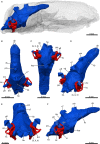
Virtual 3D‐model of the brain endocast (in blue) with reconstructed inner ear labyrinth (in red) of AMNH FARB 2227: (A) right lateral view with the skull transparent, (B) dorsal view, (C) ventral view, (D) posterodorsolateral view, (E) posterior view and (F) right lateral view. asc, anterior semicircular canal; fb, forebrain; fm, foramen magnum; hb, hindbrain; hyp, hypophysis; i.e, inner ear; lsc, lateral semicircular canal; ob, olfactory bulbs; pfl, paraflocculus; pin, pineal body; psc, posterior semicircular canal; v?, vermis?; IX, X, XI, XII, cranial nerves.
Figure 14
Figure 14
The inner ear labyrinth of AMNH FARB 2227. Virtual cast of the left inner ear labyrinth in (A) posterior, (B) lateral, and (C) dorsal views. (D,E) Horizontal cross‐sections. (F,G) Transverse μCT sections through the otic region. asc, anterior semicircular canal; bc, brain cavity; bo, basioccipital; cc, crus commune; cr, cochlear recess; d, dentary; epi, epipterygoid; fv, fenestra vestibuli; hy, hyoid; lsc, lateral semicircular canal; op, opisthotic; os, orbitosphenoid; pa, parietal; pfl, paraflocculus; pp, postparietal; pro, prootic; psc, posterior semicircular canal; q, quadrate; scc, secondary crus commune; so, supraoccipital; sq, squamosal; sur, surangular; ta, tabular; uz, unossified zone; ve, vestibule.
Figure 15
Figure 15
The inner and middle ear of AMNH FARB 2227. (A) The floor of the reconstructed braincase in ventral aspect. (B) Virtual reconstruction of the left stapes in (a) dorsal, (b), proximal, (c) distal and (d) ventral views. (C) Anterior view of the reconstructed occiput to illustrate the position of the inner ear within its bony housing. (D) Reconstructed auditory region in dorsal view. ac, anterior crus; ang, angular; art, articular; asc, anterior semicircular canal; bo, basioccipital; bpt, basipterygoid process; cf, carotid foramen; cr, cochlear recess; dpq, dorsal plate of the quadrate; epi, epipterygoid; fm, foramen magnum; fp, stapes footplate; fv, fenestra vestibuli; hy, hyoid; i.e, inner ear; jf, jugular foramen; lsc, lateral semicircular canal; op, opisthotic; pbs, parabasisphenoid; pc, posterior crus; pp, postparietal; ppf, pterygoparoccipital foramen; pra, prearticular; pro, prootic; pt, pterygoid; ptf, post‐temporal fenestra; q, quadrate; so, supraoccipital; sq, squamosal; sqd, squamosal depression; st, stapes; stf, stapedial foramen; sth, stapedial head; sur, surangular; t, trochlea; ta, tabular.
Figure 16
Figure 16
3D reconstruction of the lower jaw of AMNH FARB 2227. (A) Anterodorsolateral view of the lower jaw with the skull transparent. (B) Medial view of the right lower jaw ramus. (C) Lower jaw in dorsal view. ang, angular; art, articular; cp, coronoid process; d, dentary; PC, lower postcanine; PCs, lower postcanines; pra, prearticular; sp, splenial; sur, surangular.
Figure 17
Figure 17
Dentition and tooth replacement of AMNH FARB 2227. Three‐dimensional rendering of the right upper tooth row in (A) lateral and (B) medial view. (C,D) Virtual horizontal cross‐sections through the upper jaw. Replacement canine in red; old remnant root in yellow. Arabic numerals indicate the incisor and postcanine positions. C, upper canine; fc, functional canine; la, lacrimal; mx, maxilla; oc, old canine; pl, palatine; pmx, premaxilla; ppmx, palatal process of the maxilla; rc, replacement canine; vo, vomer.

Similar articles

Cited by

References

    1. Abdala F (1996) Los chiniquodontoideos (Synapsida, Cynodontia) sudamericanos. South American Chiniquodontoids (Synapsida, Cynodontia). PhD thesis, Facultad de Ciencias Naturales, Universidad Nacional de Tucumán.
    1. Abdala F (2007) Redescription of Platycraniellus elegans (Therapsida, Cynodontia) from the lower Triassic of South Africa, and the cladistic relationships of eutheriodonts. Palaeontology 50, 591–618.
    1. Abdala F, Allinson M (2005) The taxonomic status of Parathrinaxodon proops (Therapsida: Cynodontia), with comments on the morphology of the palate in basal cynodonts. Palaeontol Africana 41, 45–52.
    1. Abdala F, Jasinoski SC, Fernandez V (2013) Ontogeny of the early Triassic cynodont Thrinaxodon liorhinus (Therapsida): dental morphology and replacement. J Vertebr Paleontol 33, 1408–1431. - PubMed
    1. Allin EF (1975) Evolution of the mammalian middle ear. J Morphol 147, 403–437. - PubMed

Publication types

LinkOut - more resources