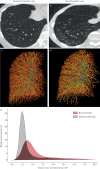
Pulmonary vascular pruning in smokers with bronchiectasis
- PMID: 30480001
- PMCID: PMC6250564
- DOI: 10.1183/23120541.00044-2018
Pulmonary vascular pruning in smokers with bronchiectasis
Abstract
There are few studies looking at the pulmonary circulation in subjects with bronchiectasis. We aimed to evaluate the intraparenchymal pulmonary vascular structure, using noncontrast chest computed tomography (CT), and its clinical implications in smokers with radiographic bronchiectasis. Visual bronchiectasis scoring and quantitative assessment of the intraparenchymal pulmonary vasculature were performed on CT scans from 486 smokers. Clinical, lung function and 6-min walk test (6MWT) data were also collected. The ratio of blood vessel volume in vessels <5 mm2 in cross-section (BV5) to total blood vessel volume (TBV) was used as measure of vascular pruning, with lower values indicating more pruning. Whole-lung and lobar BV5/TBV values were determined, and regression analyses were used to assess the differences in BV5/TBV between subjects with and without bronchiectasis. 155 (31.9%) smokers had bronchiectasis, which was, on average, mild in severity. Compared to subjects without bronchiectasis, those with lower-lobe bronchiectasis had greater vascular pruning in adjusted models. Among subjects with bronchiectasis, those with vascular pruning had lower forced expiratory volume in 1 s and 6MWT distance compared to those without vascular pruning. Smokers with mild radiographic bronchiectasis appear to have pruning of the distal pulmonary vasculature and this pruning is associated with measures of disease severity.
Conflict of interest statement
Conflict of interest: A.A. Diaz received research grants from the NIH and Brigham and Women's Hospital, and speaker fees from Novartis Inc. outside of the submitted work. Conflict of interest: D.J. Maselli has nothing to disclose. Conflict of interest: F. Rahaghi has nothing to disclose. Conflict of interest: C.E. Come received NIH/NHLBI grant K23HL114735 during the conduct of the study. Conflict of interest: A. Yen reports receiving salary support from the NIH under COPDGene R01-HL089897 during the conduct of the study. Conflict of interest: E.S. Maclean has nothing to disclose. Conflict of interest: Y. Okajima reports receiving grants from Canon Medical Systems, grants from Ziosoft, outside the submitted work. Conflict of interest: C.H. Martinez has nothing to disclose. Conflict of interest: T. Yamashiro has nothing to disclose. Conflict of interest: D.A. Lynch reports receiving grants from the NHLBI, research supports from Parexel and Veracyte, and personal fees from Boehringer Ingelheim and Genentech/Roche, outside the submitted work. Conflict of interest: W. Wang has nothing to disclose. Conflict of interest: G.L. Kinney has nothing to disclose. Conflict of interest: G.R. Washko reports receiving grants from the NIH, grants and other support from Boehringer Ingelheim, and other support from Genentech, Quantitative Imaging Solutions and PulmonX, during the conduct of the study. G.R. Washko's spouse works for Biogen, which is focused on developing therapies for fibrotic lung disease. Conflict of interest: R. San José Estépar reports receiving grants from the NHLBI during the conduct of the study. He is founder and co-owner of Quantitative Imaging Solutions, which is a company that provides image-based consulting and develops software to enable data sharing.
Figures


Similar articles
-
Pruning of the Pulmonary Vasculature in Asthma. The Severe Asthma Research Program (SARP) Cohort.Am J Respir Crit Care Med. 2018 Jul 1;198(1):39-50. doi: 10.1164/rccm.201712-2426OC. Am J Respir Crit Care Med. 2018. PMID: 29672122 Free PMC article.
-
Computed tomographic measures of pulmonary vascular morphology in smokers and their clinical implications.Am J Respir Crit Care Med. 2013 Jul 15;188(2):231-9. doi: 10.1164/rccm.201301-0162OC. Am J Respir Crit Care Med. 2013. PMID: 23656466 Free PMC article.
-
Cigarette Smoke Exposure and Radiographic Pulmonary Vascular Morphology in the Framingham Heart Study.Ann Am Thorac Soc. 2019 Jun;16(6):698-706. doi: 10.1513/AnnalsATS.201811-795OC. Ann Am Thorac Soc. 2019. PMID: 30714821 Free PMC article.
-
Vascular remodeling of the small pulmonary arteries and measures of vascular pruning on computed tomography.Pulm Circ. 2021 Nov 29;11(4):20458940211061284. doi: 10.1177/20458940211061284. eCollection 2021 Oct-Dec. Pulm Circ. 2021. PMID: 34881020 Free PMC article.
-
Quantitative computed tomography measurements to evaluate airway disease in chronic obstructive pulmonary disease: Relationship to physiological measurements, clinical index and visual assessment of airway disease.Eur J Radiol. 2016 Nov;85(11):2144-2151. doi: 10.1016/j.ejrad.2016.09.010. Epub 2016 Sep 13. Eur J Radiol. 2016. PMID: 27776670 Free PMC article. Review.
Cited by
-
Fibroblast growth factor 10 reverses cigarette smoke- and elastase-induced emphysema and pulmonary hypertension in mice.Eur Respir J. 2023 Nov 9;62(5):2201606. doi: 10.1183/13993003.01606-2022. Print 2023 Nov. Eur Respir J. 2023. PMID: 37884305 Free PMC article.
-
Suspected Bronchiectasis and Mortality in Adults With a History of Smoking Who Have Normal and Impaired Lung Function : A Cohort Study.Ann Intern Med. 2023 Oct;176(10):1340-1348. doi: 10.7326/M23-1125. Epub 2023 Oct 3. Ann Intern Med. 2023. PMID: 37782931 Free PMC article.
-
Treatment Response Evaluation by Computed Tomography Pulmonary Vasculature Analysis in Patients With Chronic Thromboembolic Pulmonary Hypertension.Korean J Radiol. 2023 Apr;24(4):349-361. doi: 10.3348/kjr.2022.0675. Epub 2023 Mar 7. Korean J Radiol. 2023. PMID: 36907594 Free PMC article.
-
Bronchial gene expression alterations associated with radiological bronchiectasis.Eur Respir J. 2023 Jan 27;61(1):2200120. doi: 10.1183/13993003.00120-2022. Print 2023 Jan. Eur Respir J. 2023. PMID: 36229050 Free PMC article.
-
Pulmonary arterial pruning is associated with CT-derived bronchiectasis progression in smokers.Respir Med. 2022 Oct;202:106971. doi: 10.1016/j.rmed.2022.106971. Epub 2022 Aug 30. Respir Med. 2022. PMID: 36116143 Free PMC article.
References
-
- Martinez-Garcia MA, de la Rosa Carrillo D, Soler-Cataluna JJ, et al. . Prognostic value of bronchiectasis in patients with moderate-to-severe chronic obstructive pulmonary disease. Am J Respir Crit Care Med 2013; 187: 823–831. - PubMed
-
- Santos S, Peinado VI, Ramirez J, et al. . Characterization of pulmonary vascular remodelling in smokers and patients with mild COPD. Eur Respir J 2002; 19: 632–638. - PubMed
Grants and funding
LinkOut - more resources
Full Text Sources

