iBet uBet web content aggregator. Adding the entire web to your favor.


Link to original content: https://pubmed.ncbi.nlm.nih.gov/29432238
Nervous System Malformations - PubMed Skip to main page content
U.S. flag

An official website of the United States government

Dot gov

The .gov means it’s official.
Federal government websites often end in .gov or .mil. Before sharing sensitive information, make sure you’re on a federal government site.

Https

The site is secure.
The https:// ensures that you are connecting to the official website and that any information you provide is encrypted and transmitted securely.

Access keys NCBI Homepage MyNCBI Homepage Main Content Main Navigation
Review
. 2018 Feb;24(1, Child Neurology):72-95.
doi: 10.1212/CON.0000000000000561.

Nervous System Malformations

Review

Nervous System Malformations

John Gaitanis et al. Continuum (Minneap Minn). 2018 Feb.

Abstract

Purpose of review: This article provides an overview of the most common nervous system malformations and serves as a reference for the latest advances in diagnosis and treatment.

Recent findings: Major advances have occurred in recognizing the genetic basis of nervous system malformations. Environmental causes of nervous system malformations, such as perinatal infections including Zika virus, are also reviewed. Treatment for nervous system malformations begins prior to birth with prevention. Folic acid supplementation reduces the risk of neural tube defects and is an important part of health maintenance for pregnant women. Fetal surgery is now available for prenatal repair of myelomeningocele and has been demonstrated to improve outcomes.

Summary: Each type of nervous system malformation is relatively uncommon, but, collectively, they constitute a large population of neurologic patients. The diagnosis of nervous system malformations begins with radiographic characterization. Genetic studies, including chromosomal microarray, targeted gene sequencing, and next-generation sequencing, are increasingly important aspects of the assessment. A genetic diagnosis may identify an associated medical condition and is necessary for family planning. Treatment consists primarily of supportive therapies for developmental delays and epilepsy, but prenatal surgery for myelomeningocele offers a glimpse of future possibilities. Prognosis depends on multiple clinical factors, including the examination findings, imaging characteristics, and genetic results. Treatment is best conducted in a multidisciplinary setting with neurology, neurosurgery, developmental pediatrics, and genetics working together as a comprehensive team.


Figures
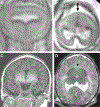
FIGURE 4–1
FIGURE 4–1
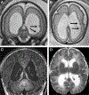
Semilobar holoprosencephaly. Coronal (A) and axial (B) fetal MRI at 20 weeks gestational age show a single ventricle, absence of the septum pellucidum, and incompletely formed interhemispheric fissure (absence of cleavage of frontal lobes [B, arrow]) consistent with holoprosencephaly. Coronal (C) and axial (D) MRI of a male newborn shows rudimental temporal and occipital lobes consistent with semilobar holoprosencephaly. Partial fusion of thalamus (D, arrowhead) and incomplete hippocampal formation are additionally recognized.
FIGURE 4–2
FIGURE 4–2
Agenesis of the corpus callosum. Fetal brain MRI (A, coronal; B, axial; C, sagittal) at 31 weeks gestational age showing complete agenesis of the corpus callosum. Characteristic findings are shown, including vertical (coronal) and parallel (axial) orientation of anterior horns of lateral ventricles and ventriculomegaly, especially of the posterior horns (colpocephaly). Full-term neonatal brain MRI (D, coronal; E, axial; F, sagittal) of the same patient with findings remnant to fetal MRI, such as parallel alignment of the bodies of lateral ventricles. Sagittal image shows abnormal radiant orientation of sulci (F). Late gestational development after 31 weeks, including gyrification, appears to have normally occurred without associated central nervous system anomalies, consistent with isolated agenesis of the corpus callosum.
FIGURE 4–3
FIGURE 4–3
Imaging and photographs of the patient in CASE 4–2. Neonatal brain MRI identifying agenesis of the corpus callosum (A, sagittal T1-weighted image), pachygyria (B, axial T2-weighted image), and parenchymal subcortical calcification (C, axial T1-weighted image). Photographs showing marked microcephaly (head circumference of 32 cm), scalloped parietal bone, slanted occiput (D, E), and diffuse spasticity (overlapping fingers) (F).
FIGURE 4–4
FIGURE 4–4
Tuberous sclerosis complex. MRI of a 4-month-old female infant with tuberous sclerosis complex. Arrowheads in T2 axial (A, B) and T1 coronal (C) images point to T2 high signal (and T1 low, not shown) cortical/subcortical (A) and cortical (B, C) tubers, and expanding gyri (A). T2 axial image (D) shows a subependymal nodule (arrow), as seen in approximately 98% of cases.
FIGURE 4–5
FIGURE 4–5
Imaging of the patient in CASE 4–3 with focal cortical dysplasia. Axial fluid-attenuated inversion recovery (FLAIR) MRI shows hyperintense signal in the left frontal cortical/subcortical region (arrow). Affected gyri appear to be wider, and their gray-white matter boundary is blurred with a thin band of T2/FLAIR hyperintensity extending from the cortex to the ventricular margin.
FIGURE 4–6
FIGURE 4–6
Periventricular nodular heterotopia. A 27-week fetus was initially referred for ventriculomegaly and found to have nodular heterotopia (A, B, arrows). Newborn MRI of this patient confirmed bilateral ventriculomegaly and periventricular nodular tissues with signal isointense to cortex suggestive for ectopic gray matter, notable in coronal (C) and axial (D) images.
FIGURE 4–7
FIGURE 4–7
Lissencephaly and TUBA1A mutation. A fetal MRI at 21 weeks gestational age was ordered because of microcephaly. Axial (A) and coronal (B) images show shallow operculum, abnormally box-shaped temporal lobes suggesting diffuse cerebral malformation. Follow-up fetal MRI at 29 weeks gestational age (C, axial; D, coronal) show persistent shallow operculum (under-undulation) and absence of normal sulcations in frontal lobes consistent with anterior dominant lissencephaly. Abnormal hypoplastic temporal lobes were also persistent. Postnatal MRI (E and F, axial; G, coronal; H, sagittal) continued to show the same features consistent with anterior dominant lissencephaly. Genetic investigation determined a TUBA1A mutation.
FIGURE 4–8
FIGURE 4–8
DCX mutation causing double cortex. Fetal MRI at 28 weeks gestational age showed smooth gyri and subcortical band heterotopia (A, axial; B, coronal; arrowheads), suggestive of lissencephaly. The image contrast was adjusted to accentuate T2 low-signal band in the subcortical region (arrowheads). Postnatal MRI (C, axial; D, coronal) of this patient confirmed subcortical band heterotopia (low-intensity signal in T2-weighted image band in subcortical white matter). The child developed global developmental delay and infantile spasms.

Similar articles

Cited by

References

    1. Barkovich AJ, Guerrini R, Kuzniecky RI, et al. A developmental and genetic classification for malformations of cortical development: update 2012. Brain 2012;135(5):1348–1369. doi:10.1093/brain/aws019. - DOI - PMC - PubMed
    1. Barkovich AJ. Subcortical heterotopia: a distinct clinicoradiologic entity. AJNR Am J Neuroradiol 1996; 17(7):1315–1322. - PMC - PubMed
    1. Barkovich AJ, Kuzniecky RI, Jackson GD, et al. Classification system for malformations of cortical development: update 2001. Neurology 2001;57(12): 2168–2178. doi:10.1212/WNL.57.12.2168. - DOI - PubMed
    1. Brody BA, Kinney HC, Kloman AS, Gilles FH. Sequence of central nervous system myelination in human infancy. I. An autopsy study of myelination. J Neuropathol Exp Neurol 1987;46(3):283–301. doi:10.1097/00005072-198705000-00005. - DOI - PubMed
    1. Greene ND, Copp AJ. Neural tube defects. Annu Rev Neurosci 2014;37:221–242. doi:10.1146/annurev-neuro-062012-170354. - DOI - PMC - PubMed