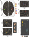
Functional architecture of the somatosensory homunculus detected by electrostimulation
- PMID: 29285773
- PMCID: PMC5830421
- DOI: 10.1113/JP275243
Functional architecture of the somatosensory homunculus detected by electrostimulation
Abstract
Key points: We performed a prospective electrostimulation study, based on 50 operated intact patients, to acquire accurate MNI coordinates of the functional areas of the somatosensory homunculus. In the contralateral BA1, the hand representation displayed not only medial-to-lateral, little-finger-to-thumb, but also rostral-to-caudal discrete somatotopy, with the tip of each finger located more caudally than the proximal phalanx. The analysis of the MNI body coordinates showed rare inter-individual variations in the medial-to-lateral somatotopic organization in these patients with rather different intensity thresholds needed to elicit sensations in different body parts. We found some similarities but also substantial differences with the previous, seminal works of Penfield and his colleagues. We propose a new drawing of the human somatosensory homunculus according to MNI space.
Abstract: In this prospective electrostimulation study, based on 50 operated patients with no sensory deficit and no brain lesion in the postcentral gyrus, we acquired coordinates in the standard MNI space of the functional areas of the somatosensory homunculus. The 3D brain volume of each patient was normalized to that space to obtain the MNI coordinates of the stimulation site locations. For 647 sites stimulated on Brodmann Area 1 (and 1025 in gyri nearby), 258 positive points for somatosensory response (40%) were found in the postcentral gyrus. In the contralateral BA1, the hand representation displayed not only medial-to-lateral and little-finger-to-thumb somatotopy, but also rostral-to-caudal discrete somatotopy, with the tip of each finger located more caudally than the proximal phalanx. We detected a medial-to-lateral, tip-to-base tongue organization but no rostral-to-caudal functional organization. The analysis of the MNI body coordinates showed rare inter-individual variations in the medial-to-lateral somatotopic organization in these patients with intact somatosensory cortex. Positive stimulations were detected through the 'on/off' outbreak effect and discriminative touch sensations were the sensations reported almost exclusively by all patients during stimulation. Mean hand (2.39 mA) and tongue (2.60 mA) positive intensity thresholds were lower (P < 0.05) than the intensities required to elicit sensations in the other parts of the body. Unlike the previous, seminal works of Penfield and colleagues, we detected no sensations such as sense of movement or desire to move, no somatosensory responses outside the postcentral gyrus, and no bilateral responses for face/tongue stimulations. We propose a rationalization of the standard drawing of the somatosensory homunculus according to MNI space.
Keywords: electrical stimulation; homunculus; somatosensory systems.
© 2017 The Authors. The Journal of Physiology © 2017 The Physiological Society.
Figures


Similar articles
-
Functional architecture of the motor homunculus detected by electrostimulation.J Physiol. 2020 Dec;598(23):5487-5504. doi: 10.1113/JP280156. Epub 2020 Sep 15. J Physiol. 2020. PMID: 32857862
-
Evidence for a rostral-to-caudal somatotopic organization in human primary somatosensory cortex with mirror-reversal in areas 3b and 1.Cereb Cortex. 2003 Sep;13(9):987-93. doi: 10.1093/cercor/13.9.987. Cereb Cortex. 2003. PMID: 12902398
-
Dermatomal Organization of SI Leg Representation in Humans: Revising the Somatosensory Homunculus.Cereb Cortex. 2017 Sep 1;27(9):4564-4569. doi: 10.1093/cercor/bhx007. Cereb Cortex. 2017. PMID: 28119344
-
Reappraisal of the somatosensory homunculus and its discontinuities.Neural Comput. 2011 Dec;23(12):3001-15. doi: 10.1162/NECO_a_00179. Neural Comput. 2011. PMID: 21732862 Review.
-
Somatotopic Mapping of the Fingers in the Somatosensory Cortex Using Functional Magnetic Resonance Imaging: A Review of Literature.Front Neuroanat. 2022 Jun 29;16:866848. doi: 10.3389/fnana.2022.866848. eCollection 2022. Front Neuroanat. 2022. PMID: 35847829 Free PMC article. Review.
Cited by
-
Ipsilateral stimulation shows somatotopy of thumb and shoulder auricular points on the left primary somatosensory cortex using high-density fNIRS.bioRxiv [Preprint]. 2024 Sep 16:2024.09.16.612477. doi: 10.1101/2024.09.16.612477. bioRxiv. 2024. PMID: 39345597 Free PMC article. Preprint.
-
Artificial embodiment displaces cortical neuromagnetic somatosensory responses.Sci Rep. 2024 Sep 27;14(1):22279. doi: 10.1038/s41598-024-72460-6. Sci Rep. 2024. PMID: 39333283 Free PMC article.
-
Evoking artificial speech perception through invasive brain stimulation for brain-computer interfaces: current challenges and future perspectives.Front Neurosci. 2024 Jun 26;18:1428256. doi: 10.3389/fnins.2024.1428256. eCollection 2024. Front Neurosci. 2024. PMID: 38988764 Free PMC article. Review.
-
Contribution of external reference frame to tactile localization.Exp Brain Res. 2024 Aug;242(8):1957-1970. doi: 10.1007/s00221-024-06877-w. Epub 2024 Jun 25. Exp Brain Res. 2024. PMID: 38918211
-
Longitudinal neurofunctional changes in medication overuse headache patients after mindfulness practice in a randomized controlled trial (the MIND-CM study).J Headache Pain. 2024 Jun 11;25(1):97. doi: 10.1186/s10194-024-01803-5. J Headache Pain. 2024. PMID: 38858629 Free PMC article. Clinical Trial.
References
-
- Blankenburg F, Meyer R, Schwiemann J & Villringer A (2003). Evidence for a rostral‐to‐caudal somatotopic organization in human primary somatosensory cortex with mirror‐reversal in areas 3b and 1. Cereb Cortex 13, 987–993. - PubMed
-
- Brodmann K (1909). Vergleichende Lokalisationslehre der Grosshirnrinde. Johann Bart, Leipzig, Germany.
-
- Darian‐Smith I, Goodwin A, Sugitani M & Heywood J (1984). The tangible features of textured surfaces: their representation in the monkey's somatosensory cortex In Dynamic Aspects of Neocortical Function, eds Edelman GM, Gall WE. & Cowan WM. Wiley & Sons, New York.
-
- Druschky K, Kaltenhäuser M, Hummel C, Druschky A, Pauli E, Huk WJ, Stefan H & Neundörfer B (2002). Somatotopic organization of the ventral and dorsal finger surface representations in human primary sensory cortex evaluated by magnetoencephalography. Neuroimage 15, 182–189. - PubMed
-
- Gardner EP (1988). Somatosensory cortical mechanisms of feature detection in tactile and kinesthetic discrimination. Can J Physiol Pharmacol 66, 439‐454. - PubMed
Publication types
MeSH terms
LinkOut - more resources
Full Text Sources
Other Literature Sources


