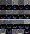
Cell reproductive patterns in the green alga Pseudokirchneriella subcapitata (=Selenastrum capricornutum) and their variations under exposure to the typical toxicants potassium dichromate and 3,5-DCP
- PMID: 28152022
- PMCID: PMC5289587
- DOI: 10.1371/journal.pone.0171259
Cell reproductive patterns in the green alga Pseudokirchneriella subcapitata (=Selenastrum capricornutum) and their variations under exposure to the typical toxicants potassium dichromate and 3,5-DCP
Abstract
Pseudokirchneriella subcapitata is a sickle-shaped freshwater green microalga that is normally found in unicellular form. Currently, it is the best known and most frequently used species of ecotoxicological bioindicator because of its high growth rate and sensitivity to toxicants. However, despite this organism's, our knowledge of its cell biology-for example, the patterns of nuclear and cytoplasmic division in the mitotic stage-is limited. Although it has been reported that P. subcapitata proliferates by popularity forming four daughter cells (autospores) through multiple fission after two nuclear divisions, here, we report two additional reproductive patterns by which two autospores are formed by binary fission ("two-autospore type") and eight autospores are formed by multiple fission ("eight-autospore type"). Moreover, we found that cell reproductive patterns differed markedly with the culture conditions or with exposure to either of two typical toxicants, potassium dichromate (K2Cr2O7) and 3,5-dichlorophenol (3,5-DCP). The eight-autospore type occurred at the highest frequency in the early phase of culture, but it disappeared under 3,5-DCP at 2.0 mg/L. Under 0.3 mg/L K2CrO7 (Cr(VI)) the eight-autospore type took substantially longer to appear than in control culture. The two-autospore type occurred only in the late phase of culture. To our knowledge, this is the first detailed evaluation of the reproductive patterns of P. subcapitata, which changed dramatically in the presence of toxicants. These findings suggest that observation of the reproductive patterns of P. subcapitata will help to elucidate different cell reactions to toxicants.
Conflict of interest statement
The authors have declared that no competing interests exist.
Figures


Similar articles
-
Features of the microalga Raphidocelis subcapitata: physiology and applications.Appl Microbiol Biotechnol. 2024 Feb 19;108(1):219. doi: 10.1007/s00253-024-13038-0. Appl Microbiol Biotechnol. 2024. PMID: 38372796 Free PMC article. Review.
-
Rapid ecotoxicological bioassay using delayed fluorescence in the green alga Pseudokirchneriella subcapitata.Water Res. 2006 Oct;40(18):3393-400. doi: 10.1016/j.watres.2006.07.016. Epub 2006 Sep 12. Water Res. 2006. PMID: 16970970
-
Modification of cell volume and proliferative capacity of Pseudokirchneriella subcapitata cells exposed to metal stress.Aquat Toxicol. 2014 Feb;147:1-6. doi: 10.1016/j.aquatox.2013.11.017. Epub 2013 Dec 4. Aquat Toxicol. 2014. PMID: 24342441
-
Reproductive cycle progression arrest and modification of cell morphology (shape and biovolume) in the alga Pseudokirchneriella subcapitata exposed to metolachlor.Aquat Toxicol. 2020 May;222:105449. doi: 10.1016/j.aquatox.2020.105449. Epub 2020 Feb 17. Aquat Toxicol. 2020. PMID: 32109756
-
Cell-cycle regulation in green algae dividing by multiple fission.J Exp Bot. 2014 Jun;65(10):2585-602. doi: 10.1093/jxb/ert466. Epub 2014 Jan 17. J Exp Bot. 2014. PMID: 24441762 Review.
Cited by
-
Features of the microalga Raphidocelis subcapitata: physiology and applications.Appl Microbiol Biotechnol. 2024 Feb 19;108(1):219. doi: 10.1007/s00253-024-13038-0. Appl Microbiol Biotechnol. 2024. PMID: 38372796 Free PMC article. Review.
-
Ecotoxicology Evaluation of a Fenton-Type Process Catalyzed with Lamellar Structures Impregnated with Fe or Cu for the Removal of Amoxicillin and Glyphosate.Int J Environ Res Public Health. 2023 Dec 13;20(24):7172. doi: 10.3390/ijerph20247172. Int J Environ Res Public Health. 2023. PMID: 38131723 Free PMC article.
-
In Silico and Cellular Differences Related to the Cell Division Process between the A and B Races of the Colonial Microalga Botryococcus braunii.Biomolecules. 2021 Oct 5;11(10):1463. doi: 10.3390/biom11101463. Biomolecules. 2021. PMID: 34680096 Free PMC article.
-
Comparative genome analysis of test algal strain NIVA-CHL1 (Raphidocelis subcapitata) maintained in microalgal culture collections worldwide.PLoS One. 2020 Nov 9;15(11):e0241889. doi: 10.1371/journal.pone.0241889. eCollection 2020. PLoS One. 2020. PMID: 33166324 Free PMC article.
-
Analyzing the toxicity of bisphenol-A to microalgae for ecotoxicological applications.Environ Monit Assess. 2019 Dec 3;192(1):8. doi: 10.1007/s10661-019-7984-0. Environ Monit Assess. 2019. PMID: 31797148
References
-
- Nygaad G, Komárek J, Kristiansen J, Skulberg OM. Taxonomic designations of the bioassay alga NIVA-CHL1 ("Selenastrum capricornum") and some related strains. Opera Botanica. 1987;90: 1–46.
-
- Hindák F. Studies on the chlorococcal algae (Chlorophyceae). V Biologické Práce. 1990;36: 1–227.
-
- Machado MD, Soares EV. Modification of cell volume and proliferative capacity of Pseudokirchneriella subcapitata cells exposed to metal stress. Aquatic Toxicol. 2014;147: 1–6. - PubMed
-
- Franklin NM, Stauber JL, Lim RP. Development of flow cytometry-based algal bioassays for assessing toxicity of copper in natural waters. Environ Toxicol Chem. 2001;20: 160–170. - PubMed
MeSH terms
Substances
Grants and funding
LinkOut - more resources
Full Text Sources
Other Literature Sources
Research Materials
Miscellaneous


