iBet uBet web content aggregator. Adding the entire web to your favor.


Link to original content: https://pubmed.ncbi.nlm.nih.gov/27894837
CD4+ virtual memory: Antigen-inexperienced T cells reside in the naïve, regulatory, and memory T cell compartments at similar frequencies, implications for autoimmunity - PubMed Skip to main page content
U.S. flag

An official website of the United States government

Dot gov

The .gov means it’s official.
Federal government websites often end in .gov or .mil. Before sharing sensitive information, make sure you’re on a federal government site.

Https

The site is secure.
The https:// ensures that you are connecting to the official website and that any information you provide is encrypted and transmitted securely.

Access keys NCBI Homepage MyNCBI Homepage Main Content Main Navigation
. 2017 Feb:77:76-88.
doi: 10.1016/j.jaut.2016.11.001. Epub 2016 Nov 25.

CD4+ virtual memory: Antigen-inexperienced T cells reside in the naïve, regulatory, and memory T cell compartments at similar frequencies, implications for autoimmunity

Affiliations

CD4+ virtual memory: Antigen-inexperienced T cells reside in the naïve, regulatory, and memory T cell compartments at similar frequencies, implications for autoimmunity

Alina I Marusina et al. J Autoimmun. 2017 Feb.

Abstract

It is widely accepted that central and effector memory CD4+ T cells originate from naïve T cells after they have encountered their cognate antigen in the setting of appropriate co-stimulation. However, if this were true the diversity of T cell receptor (TCR) sequences within the naïve T cell compartment should be far greater than that of the memory T cell compartment, which is not supported by TCR sequencing data. Here we demonstrate that aged mice with far fewer naïve T cells, respond to the model antigen, hen eggwhite lysozyme (HEL), by utilizing the same TCR sequence as their younger counterparts. CD4+ T cell repertoire analysis of highly purified T cell populations from naive animals revealed that the HEL-specific clones displayed effector and central "memory" cell surface phenotypes even prior to having encountered their cognate antigen. Furthermore, HEL-inexperienced CD4+ T cells were found to reside within the naïve, regulatory, central memory, and effector memory T cell populations at similar frequencies and the majority of the CD4+ T cells within the regulatory and memory populations were unexpanded. These findings support a new paradigm for CD4+ T cell maturation in which a specific clone can undergo a differentiation process to exhibit a "memory" or regulatory phenotype without having undergone a clonal expansion event. It also demonstrates that a foreign-specific T cell is just as likely to reside within the regulatory T cell compartment as it would the naïve compartment, arguing against the specificity of the regulatory T cell compartment being skewed towards self-reactive T cell clones. Finally, we demonstrate that the same set of foreign and autoreactive CD4+ T cell clones are repetitively generated throughout adulthood. The latter observation argues against T cell-depleting strategies or autologous stem cell transplantation as therapies for autoimmunity-as the immune system has the ability to regenerate pathogenic clones.

Keywords: Autoimmunity; CD4 T cell; Driver T cells; Experimental autoimmune encephalomyelitis; Hematopoietic stem cell transplantation; Memory T cells; Next generation sequencing; T cell repertoire analysis; T regulatory cells; Virtual memory.


Conflict of interest statement

Conflicts of Interest: The authors declare no conflict of interest.

Figures

Fig. 1.
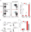
Fig. 1.. Aged and young mice expand similar sets of T cells to a HEL.
(A) Flow cytometry of mice splenocytes demonstrates that the CD4+, CD25med/low, CD44high, CD62L- T effector memory population is dramatically expanded in aged (18-month-old) BALB/c mice (p<0.001 student’s t-test), while their CD4+, CD25 med/low, CD44low, CD62L+ naïve T cell population is statistically significantly diminished (p<0.001 student’s t-test). (B) Aged (18-month-old) and young (7-week- old) BALB/c mice were immunized subcutaneously with HEL emulsified in CFA. Nine days later, draining lymph nodes were extracted and incubated with HEL or serum-free medium alone. After 72h incubation TCR CDR3 spectrotyping analysis of TCR specific for HELwas performed. Results are representative of three independent experiments. In cultures incubated with HEL, there was a considerable expansion of Vβ8.2Jβ1.5 T cells with a CDR3 length of 36 nucleotide. (C) Aged (18- month-old) and young (7-week-old) BALB/c mice were immunized subcutaneously with HEL emulsified in CFA. Lymph nodes were harvested 9 days later and primary cultures were incubated with HEL or serum-free medium alone. ELISA was performed for IFN-γ detection on the 72-hour supernatants.
Fig. 2.
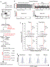
Fig. 2.. In unimmunized animals HEL-specific T cells are present in the naive, regulatory, effector memory, and central memory T cell compartments.
(A) Splenocytes from antigen-naive, 12-to-14-week-old BALB/c mice were FACS sorted to isolate naive, regulatory, effector memory, and central memory T cells using antibodies specific to CD4, CD25, CD127, CD44, and CD62L. RNA was then harvested from the isolated T cells and cDNA was synthesized using a TCR-specific primer to the constant region of the TCR β gene. To minimize amplification bias, the cDNA was then split into multiple PCR reaction tubes. Primers to Vβ8.2 and Jβ1.5 and high high-fidelity Taq DNA polymerase were then used to amplify the CDR3 region of just the Vβ8.2Jβ1.5 subpopulation of T cells. To minimize amplification bias, the number of PCR cyles was optimized by real-time PCR to ensure that PCR reactions were stopped during the linear phase of the amplification. Amplified products were used to generate sequencing libraries. 9,850,078 total reads were obtained from the central memory T cell library. These sequences were then filtered to remove sequences with incomplete CDR3 regions, N’s, and frameshifts. Sequences were also removed if they did not meet a Phred quality score cut-off of 30, or if their forward and reverse sequences did not match perfectly. The remaining 4,622,141 (central memory) CDR3 sequences were then analyzed to determine the frequency of the HEL-specific sequence. The same process was repeated for the other T cell subpopulations. Results are representative of three independent experiments. (B) The germline Vβ8.2 sequence, upstream of the CDR3 region, was used to determine the frequency of sequencing/amplification errors. Similarity scores for the different reads, and the read’s copy number are represented graphically against sequence rank order; the reads were ranked based upon their copy number with “1” being the most abundant read. Results are representative of three independent experiments. (C) Graphs of copy number vs. distinct CDR3 sequence revealed that the HEL- specific Vβ8.2Jβ1.5 CDR3 sequence was present within the naive, regulatory, effector memory, and central memory T cell populations and that the sequence was not expanded when compared with other CDR3 sequences. D96 cut off values(red dot lines) were calculated [5] to identify CDR3 sequences that resided at unacceptably low frequencies, i.e. those that had an increased possibility of originating from amplification or sequencing errors. Results are representative of three independent experiments. (D) In silico spectratyping of CDR3 lengths revealed Gaussian distributions for the naive, regulatory, central memory, and effector memory Vβ8.2Jβ1.5 spectra. Results are representative of at lest three independent experiments.
Fig 3.
Fig 3.. Frequency of the characteristic HEL-specific CD4+ T cell clone in antigen- naive and immunized mice.
(A) 4 week old BALB/c mice were injected with PBS or HEL emulsified in IFA and four day later removed by surgical excision. The mice were then sacrificed at 12 weeks and for focused Vβ8.2Jβ1.5 TCR next generation repertoire analysis was conducted on splenic T cells. Significance calculated by Student’s t-test. Graphs of copy number vs. distinct nucleotide CDR3 sequence are shown. Red arrow indicates location of HEL-specific Vβ8.2Jβ1.5 CDR3 sequence(CDR3=CASGTGNNQAPL). Results are representative of at lest three independent experiments. (B) Spleen cells from unimmunized 12 week old BALB/c mice were harvested and stained with antibodies specific to CD4, CD25, CD44, CD127, and CD62L. Lymphocytes were then FACS sorted to isolate pure populations of memory (CD4+, CD25med/low, CD44 high, CD62L−/+) T cells. Isolated memory CD4+ T cells were cocultured for 96 hr with in vitro generated BMDCs in presence or absence of HEL. RNA was extracted for focused Vβ8.2Jβ1.5 TCR next generation repertoire analysis. We were unable to generate focused Vβ8.2Jβ1 TCR libraries from BMDCs cultured alone in presence or absence of HEL. These confirms absence of T cell in our BMDCs preparations. Resulting reads were then analyzed using an in-house developed software. Graphs of copy number vs. distinct nucleotide CDR3 sequence are shown. Red arrow indicates location of HEL-specific Vβ8.2Jβ1.5 CDR3 sequence(CDR3=CASGTGNNQAPL).
Fig 4.
Fig 4.. The immune system is able to regenerate characteristic T cells after T cell depletion.
Young (7 weeks) and old (18 month) BALB/c mice were injected with 300 μg of a T cell-depleting CD4-specific monoclonal antibody. (A) One day following depletion flow cytometry was done to confirmed the selective loss of CD4+ T cells. Repeat flow cytometry 21 days later confirmed repletion of the CD4+ T cell compartment. (B) Following return of the CD4+ T cells mice were immunized subcutaneously with HEL emulsified in CFA. Lymph nodes were harvested 9 days later and primary cultures were incubated for 72h with HEL or serum-free medium alone. TCR CDR3 spectrotyping analysis revealed expansion of the characteristic HEL-specific Vβ8.2Jβ1.5 T cell. Results are representative of four independent experiments.
Fig 5.
Fig 5.. Following hematopoietic stem cell transplantation (HSCT) dominant characteristic T cells are regenerated.
(A) BALB/c mice were lethally irradiated and injected with 300 μg of a T cell- depleting CD4-specific monoclonal antibody. They were then rescued via intravenous transplantation of T cell-depleted syngeneic bone marrow cells (106 cells). Following lymphocyte recovery (6 weeks) mice were immunized subcutaneously with HEL emulsified in CFA. Lymph nodes were harvested 9 days later and primary cultures were incubated for 72h with HEL or serum-free medium alone. TCR CDR3 spectrotyping analysis reveald that the characteristic HEL-specific Vβ8.2Jβ1.5 T cell clonotype was not significantly exanded. Results are representative of four independent experiments. (B) ELISA was performed for IFN-γ detection on the 72-hour supernatants. Although the characterisitc expansion was not observed, mice were able to secrete significant amounts of IFN-γ (p = 0.007, Student’s t-test) and (C) were able to expand other HEL-specific T cell clones. (D) A second group of mice were treated exactly as described in “A” with exception that their immune system was allowed to recover for a longer period of time (100 days). Flow cytometry was done to confirmed the repletion of the CD4+ T cell compartment. Mice were immunized subcutaneously with HEL emulsified in CFA. Lymph nodes were harvested 9 days later and primary cultures were incubated for 72h with HEL or serum-free medium alone. (E) ELISA was performed for IFN-γ detection on the 72-hour supernatants. Results are representative of four independent experiments. Significant increase in IFN-γ secretion (p = 0.0014, Student’s t-test) was detected in culture supernantants when lymphocytes were incubated with HEL and (F) TCR CDR3 spectrotyping analysis demonstrated that the characteristic HEL-specific Vβ8.2Jβ1.5 T cell clonotype was strongly exanded in respone to culture with HEL. Results are representative of four independent experiments.
Fig. 6.
Fig. 6.. Following hematopoietic stem cell transplantation (HSCT) dominant autoreactive T cells are regenerated.
(A) B10.PL/J mice were lethally irradiated and injected with a CD4-depleting monoclonal antibody, GK1.5. They were then rescued via intravenous transplantation of T cell-depleted syngeneic bone marrow cells (106 cells). Flow cytometry was done to confirmed the repletion of the CD4+ T cell compartment. 100 days status-post transplantation mice were immunized with the self antigen P:Ac1–9 and lymph nodes were harvested 9 days afterwards. Non-transplanted mice were also immunized with MBP as control. Following MBP priming draining lymph nodes were harvested and incubated with either MBP or medium alone. (B) ELISA was performed for IFN-γ detection on the 72-hour supernatants. Results are representative of four independent experiments. Significant increase in IFN-γ secretion was detected in lymphocyte cultures incubated with autoantigen MBP:Ac1–9. (C) TCR CDR3 spectrotyping analysis demonstrated that the characteristic dominant Vβ8.2Jβ2.7 T cell clonotype was strongly expanded when lymphocytes were cultured with 20 mcg/ml MBP:Ac1–9. Results are representative of four independent experiments. (D) Experimental autoimmune encephalomyelitis (EAE) was induced in five mice that had undergone syngeneic HSCT and five non- transplanted control mice. Following EAE induction, mice that had undergone syngeneic HSCT developed worse EAE (p=0.01, Student’s t-test] than their none transplanted counterparts.
Fig. 7.
Fig. 7.. Pathway of conventional and unconventional naive CD4+ T cell differentiation.
Conventional view: Upon cognate antigen recognition, naive CD4+ T cells differentiate into effector cells and form “true” antigen-experienced memory cells. Proposed model: Under physiological conditions, naive CD4+ T cells may also acquire a memory or regulatory phenotype via mechanisms that do not require strong antigen recognition and T cell expansion. Driving forces for the generation of “virtual” memory T cells from naive T cells are currently unknown but may include physiologic aging of the cell, low cross-reactivity with self, and cross-recognition with commensal microbiota. The existence of “virtual” memory CD4+ T cells has direct implication for autoimmunity as it is possible for autoreactive immune responses to arise from crossreactivity directly within the T cell memory pool.

Similar articles

Cited by

References

    1. Sallusto F, Lenig D, Forster R, Lipp M, & Lanzavecchia A (1999) Two subsets of memory T lymphocytes with distinct homing potentials and effector functions. Nature 401(6754):708–712. - PubMed
    1. Bielekova B, et al. (2000) Encephalitogenic potential of the myelin basic protein peptide (amino acids 83–99) in multiple sclerosis: results of a phase II clinical trial with an altered peptide ligand. Nat Med 6(10):1167–1175. - PubMed
    1. Kimmig S, et al. (2002) Two subsets of naive T helper cells with distinct T cell receptor excision circle content in human adult peripheral blood. J Exp Med 195(6):789–794. - PMC - PubMed
    1. Davis MM & Bjorkman PJ (1988) T-cell antigen receptor genes and T-cell recognition. Nature 334(6181):395–402. - PubMed
    1. Warren RL, et al. (2011) Exhaustive T-cell repertoire sequencing of human peripheral blood samples reveals signatures of antigen selection and a directly measured repertoire size of at least 1 million clonotypes. Genome Res 21(5):790–797. - PMC - PubMed

Publication types

MeSH terms