iBet uBet web content aggregator. Adding the entire web to your favor.


Link to original content: https://pubmed.ncbi.nlm.nih.gov/27847361
Functional Characterization of Adaptive Mutations during the West African Ebola Virus Outbreak - PubMed Skip to main page content
U.S. flag

An official website of the United States government

Dot gov

The .gov means it’s official.
Federal government websites often end in .gov or .mil. Before sharing sensitive information, make sure you’re on a federal government site.

Https

The site is secure.
The https:// ensures that you are connecting to the official website and that any information you provide is encrypted and transmitted securely.

Access keys NCBI Homepage MyNCBI Homepage Main Content Main Navigation
. 2017 Jan 3;91(2):e01913-16.
doi: 10.1128/JVI.01913-16. Print 2017 Jan 15.

Functional Characterization of Adaptive Mutations during the West African Ebola Virus Outbreak

Affiliations

Functional Characterization of Adaptive Mutations during the West African Ebola Virus Outbreak

Erik Dietzel et al. J Virol. .

Abstract

The Ebola virus (EBOV) outbreak in West Africa started in December 2013, claimed more than 11,000 lives, threatened to destabilize a whole region, and showed how easily health crises can turn into humanitarian disasters. EBOV genomic sequences of the West African outbreak revealed nonsynonymous mutations, which induced considerable public attention, but their role in virus spread and disease remains obscure. In this study, we investigated the functional significance of three nonsynonymous mutations that emerged early during the West African EBOV outbreak. Almost 90% of more than 1,000 EBOV genomes sequenced during the outbreak carried the signature of three mutations: a D759G substitution in the active center of the L polymerase, an A82V substitution in the receptor binding domain of surface glycoprotein GP, and an R111C substitution in the self-assembly domain of RNA-encapsidating nucleoprotein NP. Using a newly developed virus-like particle system and reverse genetics, we found that the mutations have an impact on the functions of the respective viral proteins and on the growth of recombinant EBOVs. The mutation in L increased viral transcription and replication, whereas the mutation in NP decreased viral transcription and replication. The mutation in the receptor binding domain of the glycoprotein GP improved the efficiency of GP-mediated viral entry into target cells. Recombinant EBOVs with combinations of the three mutations showed a growth advantage over the prototype isolate Makona C7 lacking the mutations. This study showed that virus variants with improved fitness emerged early during the West African EBOV outbreak.

Importance: The dimension of the Ebola virus outbreak in West Africa was unprecedented. Amino acid substitutions in the viral L polymerase, surface glycoprotein GP, and nucleocapsid protein NP emerged, were fixed early in the outbreak, and were found in almost 90% of the sequences. Here we showed that these mutations affected the functional activity of viral proteins and improved viral growth in cell culture. Our results demonstrate emergence of adaptive changes in the Ebola virus genome during virus circulation in humans and prompt further studies on the potential role of these changes in virus transmissibility and pathogenicity.

Keywords: Ebola virus; West Africa; adaptive mutations; glycoprotein; zoonotic infections.


Figures

FIG 1
FIG 1
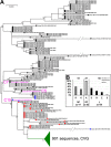
Ebola virus sequence variation at amino acid positions NP111, GP82, and L759. (A) Phylogenetic tree of the EBOV sequences obtained during the West African outbreak. The sequences are labeled with the country of sampling, the date of sampling, and the GenBank accession number. Sequences of Makona isolates C5, C7, and C15 are shown in purple font. Amino acid signatures at positions NP111, GP82, and L759 are depicted next to the sequence name. Black circles, NP111R, GP82A, and L759D (RAD = C07 signature); blue circles, NP111R, GP82A, and LD759G (RAG = point mutation in L); red circles, NP111R, GPA82V, and LD759G (RVG = point mutations in L and GP). The branch containing 910 sequences with the triple mutation NPR111C, GPA82V, and LD759G (CVG) is collapsed for clarity (green). The scale bar represents 0.0001 units of nucleotide substitutions per site. Percentages of replicate trees in which the associated taxa clustered together in more than 50% of 1,000 bootstrap replicates are shown next to the branches. (B) Sequence variations at positions NP111, GP82, and L759 in all sequenced Ebola virus genes available in the database (from 1976 to 2013) before the West African outbreak. aa, amino acids.
FIG 2
FIG 2
Emergence and fixation of three amino acid mutations at positions NP111, GP82, and L759 of Ebola Makona virus during the West African outbreak. (A) Vertical red bars in the schematic of the EBOV genome illustrate the location of the three amino acid mutations in NP, GP, and L open reading frames. Row 1, amino acid mutations at NP111, GP82, and L759; row 2, incidence of the three different mutations in 1,011 full viral genome sequences from EBOV cases that occurred between March 2014 and October 2015; row 3, date of the first appearance of mutations at positions NP111, GP82, and L759 and the respective GenBank accession numbers (in parentheses). (B) Chronological appearance of EBOV Makona mutants carrying mutations at positions NP111, GP82, and L759. From March until June, the majority of sequences had the signature of the prototype Makona C7 (69%). Sequences with the single mutation L759D were reported for a short time period (until April 2014) (see Table S1). Until end of May 2014, the double mutant GPA82V and LD75 and the triple mutant NPR111C, GPA82V, and LD759G coexisted before the triple mutation became the dominant signature in the following months until the end of the outbreak.
FIG 3
FIG 3
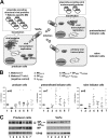
Functional analyses of the three mutations NP111, GP82, and L759 using reporter assays. (A) A schematic of the virus-like particle (VLP) assay used. HEK293 cells (producer cells) were transfected with plasmids expressing all of the viral structural proteins and a Makona-specific minigenome encoding Renilla luciferase. The minigenome is encapsidated, transcribed, and replicated by the viral nucleocapsid proteins. Viral replication and transcription are monitored by measuring Renilla luciferase activity. Replication results in the formation of mininucleocapsids which are released from cells in the form of virus-like particles (VLPs). VLPs are used to infect either naive Huh7 cells (naive indicator cells) or Huh7 cells pretransfected with plasmids encoding the EBOV nucleocapsid proteins (pretransfected indicator cells). Naive indicator cells support only primary transcription of the incoming mininucleocapsids, whereas pretransfected indicator cells support both transcription and replication of the incoming mininucleocapsids. Luc, luciferase activity; MNCs, mininucleocapsids; MG, minigenome. (B) Functional analyses of mutant EBOV proteins in a VLP assay. The VLP assay was performed by using plasmids encoding the Makona C7 structural proteins (column 1), Makona C7 plasmids without the L plasmid (column 2, negative control), and Makona C7 structural proteins with single or combinations of plasmids replaced by mutant plasmids (columns 3 to 7). The reporter gene activity was measured (using Renilla luciferase). Values were normalized to Makona C7 (value set as 1). Error bars indicate standard deviations of the results of at least seven independent experiments. P values: *, ≤0.05; **, ≤0.01. (C) Expression control of viral proteins in the cell lysates and in VLPs. Cell lysates and VLPs from the supernatants were subjected to Western blot analysis. The viral proteins were stained with monospecific antibodies against NP, VP40, and GP. Numbering of the lanes is identical to that described for panel B. GPER, G protein-coupled estrogen receptor.
FIG 4
FIG 4
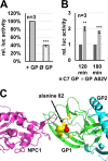
Functional analysis of GP- and GPA82V-mediated entry of VLPs. (A) Influence of GP on reporter gene activity in a VLP setting in producer cells as described in the Fig. 3A legend. Makona C7 structural proteins were expressed in Huh7 cells either with GP (+ GP) or without GP (Ø GP). Reporter gene activity was measured and normalized (presence of GP = 100%). (B) GP- and GPA82V-mediated entry of VLPs. EBOV VLPs containing a VP30/Renilla luciferase fusion protein carrying either Makona GP or Makona GPA82V were produced and purified. HEK293 cells were transduced with the purified VLPs, and the entry was monitored using the appearance of the VP30/Renilla luciferase protein in the cytosol of the transduced cells as the readout of infection (23). The reporter gene activity of cytosolic nucleocapsid-associated VP30/Renilla luciferase in the transduced cells was measured. (A and B) The error bars represent the standard deviations of the results of three independent experiments. P values: **, ≤0.01; ***, ≤0.001. (C) Location of the A82V mutation in the receptor binding domain of EBOV GP. The model is based on the X-ray structure of the GP complexed with its endosomal receptor NPC1 (Protein Data Bank [PDB] identifier 5F18). The interacting parts of GP1 and NPC1 are shown in green and purple, respectively; GP2 is shown in cyan. The red ball depicts the C-beta atom of alanine in position 82. Amino acid residues located within 4 Å of residue 82 are highlighted in yellow.
FIG 5
FIG 5
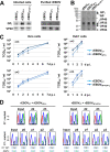
Characterization of rEBOVGP+L and rEBOVNP+GP+L. Mutations GPA82V and LD759G (rEBOVGP+L) or NPR111C, GPA82V, and LD759G (rEBOVNP+GP+L) were introduced into the genome of EBOV Makona by reverse genetics, and recombinant EBOVs were rescued. (A and B) Purified viruses were subjected to SDS-PAGE followed by Western blotting (A) and silver staining (B). Western blots were developed with chicken anti-VP40 and anti-GP antibodies and secondary IRDye680-conjugated anti-chicken antibodies. ⭑, nonviral protein band (albumin) copurified with the virions. (C) rEBOV Makona C7, rEBOVGP+L, or rEBOVNP+GP+L was used to infectVeroE6 or Huh7 cells. Samples from the supernatants were harvested at the indicated times and used to perform TCID50 assays. Error bars indicate standard deviations. d p.i., day postinfection. P values: *, ≤0.05; **, ≤0.01. (D) For the experiments whose results are depicted in the upper part of the panel, equal amounts (1:1) of PFU of rEBOV Makona and either rEBOVGP+L or rEBOVNP+GP+L were mixed and used to infect Huh7 cells. Samples were removed from the supernatants at day 2 (d2) and day 3 (d3) p.i., and viral RNA was purified. RT-PCR was performed, amplifying fragments that covered GP position 82, and amplicons were sequenced by Sanger sequencing. For the experiments whose results are depicted in the lower part of the panel, Huh7 cells were infected with a 9:1 mixture of rEBOV Makona and either rEBOVGP+L or rEBOVNP+GP+L as described above. Supernatants of infected cells were passaged at day 3 p.i. (p3) onto fresh Huh7 cells. Viral RNA was harvested and sequenced at day 3 p.i. of each passage.

Similar articles

Cited by

References

    1. Feldmann H, Geisbert TW. 2011. Ebola haemorrhagic fever. Lancet 377:849–862. doi:10.1016/S0140-6736(10)60667-8. - DOI - PMC - PubMed
    1. Bharat TA, Noda T, Riches JD, Kraehling V, Kolesnikova L, Becker S, Kawaoka Y, Briggs JA. 2012. Structural dissection of Ebola virus and its assembly determinants using cryo-electron tomography. Proc Natl Acad Sci U S A 109:4275–4280. doi:10.1073/pnas.1120453109. - DOI - PMC - PubMed
    1. Kuhn JH, Andersen KG, Baize S, Bào Y, Bavari S, Berthet N, Blinkova O, Brister JR, Clawson AN, Fair J, Gabriel M, Garry RF, Gire SK, Goba A, Gonzalez JP, Günther S, Happi CT, Jahrling PB, Kapetshi J, Kobinger G, Kugelman JR, Leroy EM, Maganga GD, Mbala PK, Moses LM, Muyembe-Tamfum JJ, N′Faly M, Nichol ST, Omilabu SA, Palacios G, Park DJ, Paweska JT, Radoshitzky SR, Rossi CA, Sabeti PC, Schieffelin JS, Schoepp RJ, Sealfon R, Swanepoel R, Towner JS, Wada J, Wauquier N, Yozwiak NL, Formenty P. 2014. Nomenclature- and database-compatible names for the two Ebola virus variants that emerged in Guinea and the Democratic Republic of the Congo in 2014. Viruses 6:4760–4799. doi:10.3390/v6114760. - DOI - PMC - PubMed
    1. WHO Ebola Response Team, Agua-Agum J, Ariyarajah A, Aylward B, Blake IM, Brennan R, Cori A, Donnelly CA, Dorigatti I, Dye C, Eckmanns T, Ferguson NM, Formenty P, Fraser C, Garcia E, Garske T, Hinsley W, Holmes D, Hugonnet S, Iyengar S, Jombart T, Krishnan R, Meijers S, Mills HL, Mohamed Y, Nedjati-Gilani G, Newton E, Nouvellet P, Pelletier L, Perkins D, Riley S, Sagrado M, Schnitzler J, Schumacher D, Shah A, Van Kerkhove MD, Varsaneux O, Wijekoon Kannangarage N. 2015. West African Ebola epidemic after one year—slowing but not yet under control. N Engl J Med 372:584–587. doi:10.1056/NEJMc1414992. - DOI - PMC - PubMed
    1. WHO. 2015. Ebola situation report. http://www.who.int/csr/disease/ebola/situation-reports/archive/en.

MeSH terms

LinkOut - more resources