Impaired consciousness in patients with absence seizures investigated by functional MRI, EEG, and behavioural measures: a cross-sectional study
- PMID: 27839650
- PMCID: PMC5504428
- DOI: 10.1016/S1474-4422(16)30295-2
Impaired consciousness in patients with absence seizures investigated by functional MRI, EEG, and behavioural measures: a cross-sectional study
Abstract
Background: The neural underpinnings of impaired consciousness and of the variable severity of behavioural deficits from one absence seizure to the next are not well understood. We aimed to measure functional MRI (fMRI) and electroencephalography (EEG) changes in absence seizures with impaired task performance compared with seizures in which performance was spared.
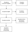
Methods: In this cross-sectional study done at the Yale School of Medicine, CT, USA, we recruited patients from 59 paediatric neurology practices in the USA. We did simultaneous EEG, fMRI, and behavioural testing in patients aged 6-19 years with childhood or juvenile absence epilepsy, and with an EEG with typical 3-4 Hz bilateral spike-wave discharges and normal background. The main outcomes were fMRI and EEG amplitudes in seizures with impaired versus spared behavioural responses analysed by t test. We also examined the timing of fMRI and EEG changes in seizures with impaired behavioural responses compared with seizures with spared responses.
Findings: 93 patients were enrolled between Jan 1, 2005, and Sept 1, 2013; we recorded 1032 seizures in 39 patients. fMRI changes during seizures occurred sequentially in three functional brain networks. In the default mode network, fMRI amplitude was 0·57% (SD 0·26) for seizures with impaired and 0·40% (0·16) for seizures with spared behavioural responses (mean difference 0·17%, 95% CI 0·11-0·23; p<0·0001). In the task-positive network, fMRI amplitude was 0·53% (SD 0·29) for seizures with impaired and 0·39% (0·15) for seizures with spared behavioral responses (mean difference 0·14%, 95% CI 0·08-0·21; p<0·0001). In the sensorimotor-thalamic network, fMRI amplitude was 0·41% (0·25) for seizures with impaired and 0·34% (0·14) for seizures with spared behavioural responses (mean difference 0·07%, 95% CI 0·01-0·13; p=0·02). Mean fractional EEG power in the frontal leads was 50·4 (SD 15·2) for seizures with impaired and 24·8 (6·5) for seizures with spared behavioural responses (mean difference 25·6, 95% CI 21·0-30·3); middle leads 35·4 (6·5) for seizures with impaired, 13·3 (3·4) for seizures with spared behavioural responses (mean difference 22·1, 95% CI 20·0-24·1); posterior leads 41·6 (5·3) for seizures with impaired, 24·6 (8·6) for seizures with spared behavioural responses (mean difference 17·0, 95% CI 14·4-19·7); p<0·0001 for all comparisons. Mean seizure duration was longer for seizures with impaired behaviour at 7·9 s (SD 6·6), compared with 3·8 s (3·0) for seizures with spared behaviour (mean difference 4·1 s, 95% CI 3·0-5·3; p<0·0001). However, larger amplitude fMRI and EEG signals occurred at the outset or even preceding seizures with behavioural impairment.
Interpretation: Impaired consciousness in absence seizures is related to the intensity of physiological changes in established networks affecting widespread regions of the brain. Increased EEG and fMRI amplitude occurs at the onset of seizures associated with behavioural impairment. These finding suggest that a vulnerable state might exist at the initiation of some absence seizures leading them to have more severe physiological changes and altered consciousness than other absence seizures.
Funding: National Institutes of Health, National Institute of Neurological Disorders and Stroke, National Center for Advancing Translational Science, the Loughridge Williams Foundation, and the Betsy and Jonathan Blattmachr Family.
Copyright © 2016 Elsevier Ltd. All rights reserved.
Figures


Comment in
-
Searching for the mechanisms of consciousness in epilepsy.Lancet Neurol. 2016 Dec;15(13):1298-1299. doi: 10.1016/S1474-4422(16)30278-2. Lancet Neurol. 2016. PMID: 27839635 Free PMC article. No abstract available.
Similar articles
-
Simultaneous EEG, fMRI, and behavior in typical childhood absence seizures.Epilepsia. 2010 Oct;51(10):2011-22. doi: 10.1111/j.1528-1167.2010.02652.x. Epilepsia. 2010. PMID: 20608963 Free PMC article.
-
Dynamic time course of typical childhood absence seizures: EEG, behavior, and functional magnetic resonance imaging.J Neurosci. 2010 Apr 28;30(17):5884-93. doi: 10.1523/JNEUROSCI.5101-09.2010. J Neurosci. 2010. PMID: 20427649 Free PMC article.
-
EEG-fMRI study on the interictal and ictal generalized spike-wave discharges in patients with childhood absence epilepsy.Epilepsy Res. 2009 Dec;87(2-3):160-8. doi: 10.1016/j.eplepsyres.2009.08.018. Epilepsy Res. 2009. PMID: 19836209
-
Finding a way in: a review and practical evaluation of fMRI and EEG for detection and assessment in disorders of consciousness.Neurosci Biobehav Rev. 2013 Sep;37(8):1403-19. doi: 10.1016/j.neubiorev.2013.05.004. Epub 2013 May 13. Neurosci Biobehav Rev. 2013. PMID: 23680699 Review.
-
Consciousness and epilepsy: why are patients with absence seizures absent?Prog Brain Res. 2005;150:271-86. doi: 10.1016/S0079-6123(05)50020-7. Prog Brain Res. 2005. PMID: 16186030 Free PMC article. Review.
Cited by
-
A human brain network linked to restoration of consciousness after deep brain stimulation.medRxiv [Preprint]. 2024 Oct 18:2024.10.17.24314458. doi: 10.1101/2024.10.17.24314458. medRxiv. 2024. PMID: 39484242 Free PMC article. Preprint.
-
Shared subcortical arousal systems across sensory modalities during transient modulation of attention.bioRxiv [Preprint]. 2024 Oct 1:2024.09.16.613316. doi: 10.1101/2024.09.16.613316. bioRxiv. 2024. PMID: 39345640 Free PMC article. Preprint.
-
Molecular Mechanisms Underlying the Generation of Absence Seizures: Identification of Potential Targets for Therapeutic Intervention.Int J Mol Sci. 2024 Sep 11;25(18):9821. doi: 10.3390/ijms25189821. Int J Mol Sci. 2024. PMID: 39337309 Free PMC article. Review.
-
The epileptic blip syndrome.Epilepsy Behav Rep. 2024 Jun 26;27:100691. doi: 10.1016/j.ebr.2024.100691. eCollection 2024. Epilepsy Behav Rep. 2024. PMID: 39050405 Free PMC article.
-
Content-state dimensions characterize different types of neuronal markers of consciousness.Neurosci Conscious. 2024 Jul 12;2024(1):niae027. doi: 10.1093/nc/niae027. eCollection 2024. Neurosci Conscious. 2024. PMID: 39011546 Free PMC article.
References
-
- Tononi G. Consciousness as integrated information: a provisional manifesto. Biol Bull. 2008;215(3):216–42. - PubMed
-
- Koch C. The quest for consciousness: a neurobiological approach. Denver, Colo.: Roberts and Co; 2004.
-
- Dehaene S. Consciousness and the brain: Deciphering how the brain codes our thoughts. New York: Viking Penguin; 2014.
-
- Laureys S, Gosseries O, Tononi G. The Neurology of Consciousness: Cognitive Neuroscience and Neuropathology. 2nd. Academic Press; 2015.
-
- Posner JB, Saper CB, Schiff ND, Plum F. Plum and Posner's Diagnosis of Stupor and Coma. 4th. Oxford University Press; USA: 2007.
MeSH terms
Grants and funding
LinkOut - more resources
Full Text Sources
Other Literature Sources


