Two New Cynodonts (Therapsida) from the Middle-Early Late Triassic of Brazil and Comments on South American Probainognathians
- PMID: 27706191
- PMCID: PMC5051967
- DOI: 10.1371/journal.pone.0162945
Two New Cynodonts (Therapsida) from the Middle-Early Late Triassic of Brazil and Comments on South American Probainognathians
Abstract
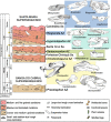
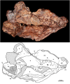
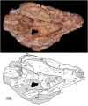
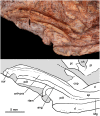
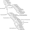
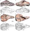
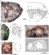
We describe two new cynodonts from the early Late Triassic of southern Brazil. One taxon, Bonacynodon schultzi gen. et sp. nov., comes from the lower Carnian Dinodontosaurus AZ, being correlated with the faunal association at the upper half of the lower member of the Chañares Formation (Ischigualasto-Villa Unión Basin, Argentina). Phylogenetically, Bonacynodon is a closer relative to Probainognathus jenseni than to any other probainognathian, bearing conspicuous canines with a denticulate distal margin. The other new taxon is Santacruzgnathus abdalai gen. et sp. nov. from the Carnian Santacruzodon AZ. Although based exclusively on a partial lower jaw, it represents a probainognathian close to Prozostrodon from the Hyperodapedon AZ and to Brasilodon, Brasilitherium and Botucaraitherium from the Riograndia AZ. The two new cynodonts and the phylogenetic hypothesis presented herein indicate the degree to which our knowledge on probainognathian cynodonts is incomplete and also the relevance of the South American fossil record for understanding their evolutionary significance. The taxonomic diversity and abundance of probainognathians from Brazil and Argentina will form the basis of deep and complex studies to address the evolutionary transformations of cynodonts leading to mammals.
Conflict of interest statement
The authors have declared that no competing interests exist.
Figures







Similar articles
-
A new prozostrodontian cynodont (Therapsida) from the Late Triassic Riograndia Assemblage Zone (Santa Maria Supersequence) of Southern Brazil.An Acad Bras Cienc. 2014 Dec;86(4):1673-92. doi: 10.1590/0001-3765201420140455. Epub 2014 Nov 28. An Acad Bras Cienc. 2014. PMID: 25590707
-
A new cynodont from the Upper Triassic Los Colorados Formation (Argentina, South America) reveals a novel paleobiogeographic context for mammalian ancestors.Sci Rep. 2022 Apr 25;12(1):6451. doi: 10.1038/s41598-022-10486-4. Sci Rep. 2022. PMID: 35468982 Free PMC article.
-
A new early-diverging probainognathian cynodont and a revision of the occurrence of cf. Aleodon from the Chañares Formation, northwestern Argentina: New clues on the faunistic composition of the latest Middle-?earliest Late Triassic Tarjadia Assemblage Zone.Anat Rec (Hoboken). 2024 Apr;307(4):818-850. doi: 10.1002/ar.25388. Epub 2024 Jan 29. Anat Rec (Hoboken). 2024. PMID: 38282519
-
A new fossil from the Jurassic of Patagonia reveals the early basicranial evolution and the origins of Crocodyliformes.Biol Rev Camb Philos Soc. 2013 Nov;88(4):862-72. doi: 10.1111/brv.12030. Epub 2013 Feb 28. Biol Rev Camb Philos Soc. 2013. PMID: 23445256 Review.
-
The origin and early evolution of dinosaurs.Biol Rev Camb Philos Soc. 2010 Feb;85(1):55-110. doi: 10.1111/j.1469-185X.2009.00094.x. Epub 2009 Nov 6. Biol Rev Camb Philos Soc. 2010. PMID: 19895605 Review.
Cited by
-
Brazilian fossils reveal homoplasy in the oldest mammalian jaw joint.Nature. 2024 Oct;634(8033):381-388. doi: 10.1038/s41586-024-07971-3. Epub 2024 Sep 25. Nature. 2024. PMID: 39322670 Free PMC article.
-
The earliest-known mammaliaform fossil from Greenland sheds light on origin of mammals.Proc Natl Acad Sci U S A. 2020 Oct 27;117(43):26861-26867. doi: 10.1073/pnas.2012437117. Epub 2020 Oct 12. Proc Natl Acad Sci U S A. 2020. PMID: 33046636 Free PMC article.
-
First record of a basal mammaliamorph from the early Late Triassic Ischigualasto Formation of Argentina.PLoS One. 2019 Aug 7;14(8):e0218791. doi: 10.1371/journal.pone.0218791. eCollection 2019. PLoS One. 2019. PMID: 31390368 Free PMC article.
-
Early evidence of molariform hypsodonty in a Triassic stem-mammal.Nat Commun. 2019 Jun 28;10(1):2841. doi: 10.1038/s41467-019-10719-7. Nat Commun. 2019. PMID: 31253810 Free PMC article.
-
A proposed terminology for the dentition of gomphodont cynodonts and dental morphology in Diademodontidae and Trirachodontidae.PeerJ. 2019 Jun 13;7:e6752. doi: 10.7717/peerj.6752. eCollection 2019. PeerJ. 2019. PMID: 31223521 Free PMC article.
References
-
- Bonaparte JF. Annotated list of the South American Triassic tetrapods In: Haughton SH, editors. Second Gondwana Symposium, Proceedings and Papers (South Africa 1970). Pretoria: CSIR; 1970. pp. 665–682.
-
- Kokogian D, Spalletti LA, Morel EM, Artabe AE, Martínez RN, Alcober OA, et al. Estratigrafía del Triásico argentino In: Artabe AE, Morel EM, Zamuner AB, editors. El Sistema Triásico en la Argentina. La Plata: Fundación Museo de La Plata Francisco Pascasio Moreno; 2001. pp. 23–54.
-
- Marsicano C, Gallego O, Arcucci A. Faunas del Triásico: relaciones, patrones de distribucion y sucesión temporal In: Artabe AE, Morel EM, Zamuner AB, editors. El Sistema Triásico en la Argentina. La Plata: Fundación Museo de La Plata “Francisco Pascasio Moreno”; 2001. pp. 131–141.
-
- Langer MC, Ribeiro AM, Schultz CL, Ferigolo J. The continental tetrapod bearing Triassic of south Brazil. Bulletin of the New Mexico Museum of Natural History and Science. 2007; 41:201–218.
-
- Abdala F, Ribeiro MA. Distribution and diversity patterns of Triassic cynodonts (Therapsida, Cynodontia) in Gondwana. Palaeogeogr Palaeoclimatol Palaeoecol. 2010; 286:202–217.
MeSH terms
Grants and funding
LinkOut - more resources
Full Text Sources
Other Literature Sources


