iBet uBet web content aggregator. Adding the entire web to your favor.


Link to original content: https://pubmed.ncbi.nlm.nih.gov/27651315
BRD4 Regulates Breast Cancer Dissemination through Jagged1/Notch1 Signaling - PubMed Skip to main page content
U.S. flag

An official website of the United States government

Dot gov

The .gov means it’s official.
Federal government websites often end in .gov or .mil. Before sharing sensitive information, make sure you’re on a federal government site.

Https

The site is secure.
The https:// ensures that you are connecting to the official website and that any information you provide is encrypted and transmitted securely.

Access keys NCBI Homepage MyNCBI Homepage Main Content Main Navigation
. 2016 Nov 15;76(22):6555-6567.
doi: 10.1158/0008-5472.CAN-16-0559. Epub 2016 Sep 20.

BRD4 Regulates Breast Cancer Dissemination through Jagged1/Notch1 Signaling

Affiliations

BRD4 Regulates Breast Cancer Dissemination through Jagged1/Notch1 Signaling

Guillaume Andrieu et al. Cancer Res. .

Abstract

The bromodomain and extraterminal (BET) proteins are epigenetic "readers" of acetylated histones in chromatin and have been identified as promising therapeutic targets in diverse cancers. However, it remains unclear how individual family members participate in cancer progression and small molecule inhibitors such as JQ1 can target functionally independent BET proteins. Here, we report a signaling pathway involving BRD4 and the ligand/receptor pair Jagged1/Notch1 that sustains triple-negative breast cancer migration and invasion. BRD4, but not BRD2 or BRD3, regulated Jagged1 expression and Notch1 signaling. BRD4-selective knockdown suppressed Notch1 activity and impeded breast cancer migration and invasion. BRD4 was required for IL6-stimulated, Notch1-induced migration and invasion, coupling microenvironment inflammation with cancer propagation. Moreover, in patients, BRD4 and Jagged1 expression positively correlated with the presence of distant metastases. These results identify a BRD4/Jagged1/Notch1 signaling pathway that is critical for dissemination of triple-negative breast cancer. Cancer Res; 76(22); 6555-67. ©2016 AACR.


Figures

Figure 1
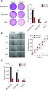
Figure 1. BET protein inhibition reduces breast cancer cell migration and invasion
(A) MDA-MB-231, SUM149PT and MCF-7 cells were pre-treated with either 400 nM (R)-JQ1 or (S)-JQ1 for 3 h then challenged for migration for 6 h with 10% FBS, using a Transwell system. Results are presented as percentage of migrated area in comparison to total membrane area. Left panel: representative photographs of the total membrane area showing migrated cells stained with crystal violet. Right panel: bars show means ± SEM of four independent experiments. Statistical analyses were performed by using Student's t test. (B) MDA-MB-231 confluent monolayer cell cultures were pre-treated with either 400 nM (R)-JQ1 or (S)-JQ1 for 3 h, then scratched with a pipet tip. Photographs were taken every two hours and the percentage of the scratch closure was determined. Left panel: representative photographs of scratched monolayer cultures of MDA-MB-231 cells treated with either (R)-JQ1 or (S)-JQ1 at different times. Right panel: means ± SEM of two independent experiments were plotted. Statistical analyses were performed by using two-way ANOVA. (C) MDA-MB-231, SUM149PT and MCF-7 cells were pre-treated with either 400 nM (R)-JQ1 or (S)-JQ1 for 3 h under conditions of serum deprivation, then plated onto Matrigel in Transwell inserts and challenged for invasion for 16 h with 10% FBS. Results are presented as the total number of invading cells. Bars show means ± SEM of four independent experiments. Statistical analyses were performed by using Student's t test. The following symbols were used to indicate significant differences: ns, p > 0.05; *, p < 0.05; **, p < 0.01; ***, p < 0.001.
Figure 2
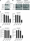
Figure 2. BRD4 regulates breast cancer cell migration and invasion
(A) Endogenous expression of BET proteins in several breast cancer cell lines was detected by immunoblotting. Molecular weights (MW) in kDa corresponding to the immunoblotted proteins are indicated. (B) siRNA-mediated BET protein depletion was validated by immunoblotting. Cells were transfected for 72h with the indicated siRNAs (50 nM). Blots shown are representative of three independent experiments. (C-D) MDA-MB-231 (left panel) and SUM149PT (right panel) cells were transfected with either control siRNA (scramble) or BET-targeted siRNAs for 72h prior to migration (C) or invasion (D) assays. Bars show means ± SEM of three independent experiments. Statistical analyses were performed by using two-way ANOVA. The following symbols were used to indicate significant differences: ns, p > 0.05; ***, p < 0.001.
Figure 3
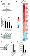
Figure 3. Identification of JAG1 as a BRD4-dependent gene
(A) Screening strategy. (B) Heatmap representing relative expression of 46 genes involved in regulation of migration and invasion in response to BRD4 depletion in MDA-MB-231 cells. Z scores are represented using a color code. Results for separate knockdown experiments are shown (n = 3). (C-D) Jagged1 mRNA (C) and protein (D) expression were measured in MDA-MB-231 cells 72h after transfection with the indicated siRNAs (50 nM). (E) Immunoblot validates Jagged1 depletion in MDA-MB-231 48h after transfection with the indicated siRNAs (20 nM) (left panel). Migration assay was conducted with scramble or siJagged1-depleted MDA-MB-231 cells (right panel). Bars show means ± SEM of three independent experiments. Statistical analyses were performed by using Student's t test. (F) Invasion assay was conducted with scrambled siRNA- or siJagged1-depleted MDA-MB-231 cells. Bars show means ± SEM of three independent experiments. Statistical analyses were performed by using Student's t test. The following symbols were used to indicate significant differences: ns, p > 0.05; **, p < 0.01; ***, p < 0.001
Figure 4
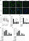
Figure 4. Jagged1/Notch1 signaling pathway acts downstream of BRD4 to regulate breast cancer cell migration and invasion
(A) Representative images from immunofluorescence experiments showing the cellular NICD1 distribution. NICD1 is stained in green and nuclei in blue, with DAPI in merged images (lower panels). Scale bar: 50 μm. (B) NICD1 nuclear score (for calculation details, see Experimental Procedures section). One dot represents a measurement of one cell. Bars show means ± SEM of three independent experiments. Statistical analyses were performed by using one-way ANOVA. (C-D) MDA-MB-231 were transfected with the indicated siRNAs (scramble, siBRD4: 50 nM; siJagged1: 20 nM) for 72h prior to conduct migration (C) and invasion (D) assays. Bars show means ± SEM of three independent experiments. Statistical analyses were performed by using one-way ANOVA. (E-F) MDA-MB-231 were transfected with the indicated siRNAs (scramble, siBRD4: 50 nM; siJagged1: 20 nM) for 72h and plasmids for 24h prior to conduct migration (E) and invasion (F) assays. Bars show means ± SEM of three independent experiments. Statistical analyses were performed by using one-way ANOVA. The following symbols were used to indicate significant differences: ns, p > 0.05; *, p < 0.05; **, p < 0.01; ***, p < 0.001.
Figure 5
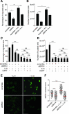
Figure 5. IL-6 induces BRD4-dependent Notch1 signaling to regulate breast cancer aggressive properties
(A-B) MDA-MB-231 cells were treated with recombinant human IL-6 (50 ng/ml) for 24h under either siBRD4 or siRNA scramble control conditions, to conduct migration (A) and invasion (B) assays. Bars show means ± SEM of three independent experiments. Statistical analyses were performed with one-way ANOVA. (C-D) MDA-MB-231 cells were treated with recombinant human IL-6 (50 ng/ml, 24h) and/or with DAPT (10 μM, 16h) under either siBRD4 or siRNA scramble control conditions, to conduct migration (C) and invasion (D) assays. Bars show means ± SEM of three independent experiments. Statistical analyses were performed with one-way ANOVA. (E) Representative images from immunofluorescence experiments showing cellular NICD1 distribution. NICD1 is stained in green and nuclei in blue with DAPI in merged images (lower panels). Scale bar: 50 μm. (F) NICD1 nuclear score. One dot represents a measurement of one cell. Statistical analyses of three independent experiments were performed with one-way ANOVA. The following symbols were used to indicate significant differences: ns, p > 0.05; *, p < 0.05; **, p < 0.01; ***, p < 0.001.
Figure 6
Figure 6. BRD4 interacts with the JAG1 promoter
(A) Immunoblots of BRD4, Jagged1, phospho-STAT3 (p-STAT3) and STAT3 in MDA-MB-231 cells treated or not with IL-6 (50 μg/ml, 24h) under either siBRD4 or siRNA scramble control conditions. Quantifications relative to control with normalization using α-tubulin levels as a loading control are indicated. The ration p-STAT3/STAT3 is also indicated to illustrate STAT3 activation. (B) Immunoblots of BRD4 and Jagged1 in MDA-MB-231 or MCF-7 cells under overexpression of the BET proteins for 48h. Quantifications relative to control with normalization using α-tubulin levels as a loading control are indicated. (C-D) MDA-MB-231 cells were treated with either 400 nM (R)- or (S)-JQ1 for 24h as indicated then harvested for ChIP. (C) BRD4 interacts with the proximal JAG1 promoter but is absent from its distal promoter. (D) BRD4 is enriched at the proximal JAG1 promoter under IL-6 treatment (50 ng/ml for 24h). The following symbols were used to indicate significant differences: ns, p > 0.05; **, p < 0.01; ***, p < 0.001.
Figure 7
Figure 7. Proposed model: a BRD4/Jagged1/Notch1 signaling pathway to regulate breast cancer dissemination
Breast cancer microenvironment is composed of diverse cell types including notably adipocytes, fibroblasts or immune cells, secreting numerous chemoattractant factors such as growth factors, chemokines or cytokines, including IL-6, that sustain cell migration or invasion. We identified BRD4 as a regulator of Jagged1, a Notch1 ligand, which regulates Notch1 signaling activity in breast cancer cells to modulate their migration and invasion. The pro-inflammatory cytokine IL-6 enriches BRD4 at the Jagged 1 promoter to stimulate its expression and promote breast cancer cell migration and invasion through Notch1 signaling. A combined elevated expression of BRD4 and Jagged1 in breast cancer associates in vivo with the presence of distant metastases.

Similar articles

Cited by

References

    1. Wu SY, Chiang CM. The double bromodomain-containing chromatin adaptor Brd4 and transcriptional regulation. J Biol Chem. 2007;282(18):13141–5. - PubMed
    1. Belkina AC, Denis GV. BET domain co-regulators in obesity, inflammation and cancer. Nature reviews Cancer. 2012;12(7):465–77. - PMC - PubMed
    1. Denis GV, Green MR. A novel, mitogen-activated nuclear kinase is related to a Drosophila developmental regulator. Genes Dev. 1996;10(3):261–71. - PubMed
    1. Denis GV, Vaziri C, Guo N, Faller DV. RING3 kinase transactivates promoters of cell cycle regulatory genes through E2F. Cell Growth Differ. 2000;11(8):417–24. - PMC - PubMed
    1. Guo N, Faller DV, Denis GV. Activation-induced nuclear translocation of RING3. J Cell Sci. 2000;113(Pt 17):3085–91. - PMC - PubMed