iBet uBet web content aggregator. Adding the entire web to your favor.


Link to original content: https://pubmed.ncbi.nlm.nih.gov/27441572
Development and Antiparkinsonian Activity of VU0418506, a Selective Positive Allosteric Modulator of Metabotropic Glutamate Receptor 4 Homomers without Activity at mGlu2/4 Heteromers - PubMed Skip to main page content
U.S. flag

An official website of the United States government

Dot gov

The .gov means it’s official.
Federal government websites often end in .gov or .mil. Before sharing sensitive information, make sure you’re on a federal government site.

Https

The site is secure.
The https:// ensures that you are connecting to the official website and that any information you provide is encrypted and transmitted securely.

Access keys NCBI Homepage MyNCBI Homepage Main Content Main Navigation
. 2016 Sep 21;7(9):1201-11.
doi: 10.1021/acschemneuro.6b00036. Epub 2016 Aug 5.

Development and Antiparkinsonian Activity of VU0418506, a Selective Positive Allosteric Modulator of Metabotropic Glutamate Receptor 4 Homomers without Activity at mGlu2/4 Heteromers

Affiliations

Development and Antiparkinsonian Activity of VU0418506, a Selective Positive Allosteric Modulator of Metabotropic Glutamate Receptor 4 Homomers without Activity at mGlu2/4 Heteromers

Colleen M Niswender et al. ACS Chem Neurosci. .

Abstract

Metabotropic glutamate receptor 4 (mGlu4) is emerging as a potential therapeutic target for numerous central nervous system indications, including Parkinson's disease (PD). As the glutamate binding sites among the eight mGlu receptors are highly conserved, modulation of receptor activity via allosteric sites within the receptor transmembrane domains using positive and negative allosteric modulators (PAMs and NAMs, respectively) has become a common strategy. We and others have used PAMs targeting mGlu4 to show that potentiation of receptor signaling induces antiparkinsonian activity in a variety of PD animal models, including haloperidol-induced catalepsy and 6-hydroxydopamine-induced lesion. Recently, mGlu4 has been reported to form heteromeric complexes with other mGlu receptor subtypes, such as mGlu2, and the resulting heteromer exhibits a distinct pharmacological profile in response to allosteric modulators. For example, some mGlu4 PAMs do not appear to potentiate glutamate activity when mGlu2 and mGlu4 are coexpressed, whereas other compounds potentiate mGlu4 responses regardless of mGlu2 coexpression. We report here the discovery and characterization of VU0418506, a novel mGlu4 PAM with activity in rodent PD models. Using pharmacological approaches and Complemented Donor-Acceptor resonance energy transfer (CODA-RET) technology, we find that VU0418506 does not potentiate agonist-induced activity when mGlu2 and mGlu4 are heterodimerized, suggesting that the antiparkinsonian action of mGlu4 PAMs can be induced by compounds without activity at mGlu2/4 heteromers.

Keywords: Allosteric modulator; Parkinson’s disease; metabotropic glutamate receptor.


Figures

Figure 1
Figure 1
In vitro pharmacological profile of VU0418506, a potent mGlu4 PAM. (A) Structure of VU0418506. (B) Calcium traces for a representative experiment performed in the presence and absence of 30 µM VU0418506. In this “triple addition” assay, a baseline read is taken for 2 s and compound or vehicle is added (designated with arrow and “compound add”). After approximately 2 min, a concentration of glutamate that elicits a 20% maximal response (“EC20 add”) is added. This addition detects the activity of potentiators. In the final addition (“EC80 add”), a concentration of glutamate that elicits an EC80 response is added. In this chimeric G protein assay, we anticipate that glutamate does not induce full coupling; inclusion of a PAM further stabilizes the active receptor state, resulting in a response in the presence of the PAM that is greater than the maximal response elicited by a saturating concentration of glutamate. As shown in the “compound add” section of the trace, VU0418506 does not elicit activity in this assay in the absence of glutamate. (C) Potency of VU0418506 in the absence (black symbols) and presence (white symbols) of an EC20 concentration of glutamate at human mGlu4 using the Gqi5-mediated calcium assay reflected in panel (B). The pEC50 is 7.24 ± 0.04 (mean ± SEM), EC50 = 59.6 nM, N = 10 determinations performed in triplicate. (D) Potency of rat and human mGlu4 using native coupling to GIRK channels as a readout. For rat mGlu4, the pEC50 is 7.34 ± 0.04 (mean ± SEM), EC50 = 46.6 nM, N = 3 determinations performed in triplicate. For human mGlu4, the pEC50 is 7.27 ± 0.08 (mean ± SEM), EC50 = 55.7 nM, N = 3 determinations performed in singlicate. (E, F) Increasing concentrations of VU0418506 were added to human mGlu4/Gqi5 cells or rat mGlu4/GIRK cells approximately 2 min prior to the addition of increasing concentrations of glutamate. Data represent three independent determinations performed in duplicate.
Figure 2
Figure 2
VU0418506 exhibits antiparkinsonian activity in reversing haloperidol-induced catalepsy at unbound brain and CSF concentrations at or above the in vitro EC50 at rat mGlu4. (A) A 1.5 mg/kg dose of haloperidol was given to rats, followed 1 h later by increasing doses of VU0418506 given orally; after 30 min, catalepsy was assessed. 56.6 mg/kg, dosed orally, of the adenosine A2A antagonist compound 23 (gray bar) was used as a positive control. *p < 0.0001, one-way ANOVA with Bonferroni post-test. Data represent mean ± SEM, N = 9–10 animals. (B) Total (black circles) and predicted unbound/free (white circles) brain levels were determined. Calculated EC50 values were 7.1 µM (total) and 34 nM (unbound/free). Data represent mean ± SEM, N = 9–10 animals. (C) CSF levels of VU0418506 were determined after oral dosing and correlated with HIC reversal. Statistically significant reversals in HIC (data from panel (A)) were observed when CSF concentrations exceeded in the in vitro EC50 at rat mGlu4. (D) Concentration–response curve of CSF concentration versus response, with a calculated EC50 value of 210 nM.
Figure 3
Figure 3
VU0418506 exhibits antiparksonian activity out to 6 h after administration, paralleling exposure levels. (A) Animals were administered vehicle or a 30 mg/kg dose of VU0418506 and efficacy in reversing catalepsy was assessed 0.5, 1, 2, 4, or 6 h after dosing. Haloperidol was given 1.5 h before each catalepsy measurement as described in Methods. **p < 0.01, ***p < 0.0001. Data represent mean ± SEM, N = 8–10 animals. (B) Plasma (white squares) and brain (black squares) exposures were calculated up to 6 h after a 30 mg/kg dose of VU0418506. Data represent the average calculated from two animals.
Figure 4
Figure 4
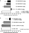
VU0418506 reverses forelimb asymmetry deficits, both alone and with a subthreshold dose of L-DOPA, in a 6-OHDA model of Parkinson’s disease. (A) 2.5 mg/kg dose of L-DOPA (hatched bar), as well as 10 and 30 mg/kg VU0418506 (gray and black bars, respectively), significantly reverse forelimb asymmetry deficits (p-values shown in figure, one-way ANOVA with Dunnett’s multiple comparison test). (B) Doses of 0.75 mg/kg L-DOPA and 3 mg/kg VU0418506 are ineffective alone, but, when paired together, significant reversals of forelimb asymmetry phenotypes are observed (*p < 0.0001 versus vehicle, one-way ANOVA with Dunnett’s multiple comparison test). Dose groups for both studies represent mean ± SEM from 6 to 18 animals.
Figure 5
Figure 5
Differential interactions of (A) VU0418506 and (B) Lu AF21934 with mGlu4 in the presence and absence of mGlu2. Increasing concentrations of compound were applied prior to the application of increasing concentrations of glutamate and concentration-response curves were assessed. Data represent three independent experiments performed in duplicate.
Figure 6
Figure 6
Schematic of CODA-RET technology to detect activity of heteromeric mGlu2/4. (A) When expressed alone, mGlu4 or mGlu2 (mGlu4 shown) form obligate homodimers due to disulfide linkages. If only mGlu4 receptors labeled with the L1 fragment of luciferase are expressed, no complementation between homomers occurs and no luminescence or BRET signal is detected. (B) If mGlu4 receptors tagged with L1 and L2 are coexpressed, luciferase is complemented and a BRET signal is produced upon G protein recruitment. (C) If mGlu4 tagged with L1 and mGlu2 tagged with L2 are expressed, complementation occurs, resulting in a BRET signal with G protein. This allows detection of a signal from heteromer activation without contamination of the signal by homomeric mGlu2 or mGlu4.
Figure 7
Figure 7
CODA-RET technology reveals that VU0418506 is ineffective in the mGlu2/4 heteromeric conformation while Lu AF21934 induces potentiation. (A, C) As illustrated in Figure 6, receptor constructs were generated in which mGlu4 was labeled with either L1 or L2 of Renilla luciferase as described in Methods and expressed in cells with mVenus-labeled Gαi G protein as well as unlabeled Gβ1 and Gγ2. Both VU0418506 and Lu AF21934 were able to potentiate responses by shifting the curve to the left ((A) pEC50 DMSO, 6.75 ± 0.07, mean ± SEM, pEC50 VU0418506, 7.91 ± , 0.06, *p = 0.0039; max response DMSO 0.029 ± 0.001, max response VU0418506, 0.047 ± 0.0004, *p = 0.0005; (C) pEC50 DMSO, 6.75 ± 0.07, pEC50 Lu AF21934, 7.70 ± 0.01, *p = 0.0026; max response DMSO, 0.029 ± 0.001, max response Lu AF21934, 0.050 ± 0.003, *p = 0.0043). (B, D) In contrast, in cells coexpressing labeled mGlu2 and mGlu4 with mVenus-labeled Gai G protein, VU0418506 was unable to potentiate responses, while Lu AF21934 induced potentiation ((B) pEC50 DMSO, 7.27 ± 0.19, mean ± SEM, pEC50 VU0418506, 7.31 ± , 0.17, *p = 0.274; max response DMSO, 0.021 ± 0.002, max response VU0418506, 0.021 ± 0.002, *p = 0.196; (D) pEC50 DMSO, 7.27 ± 0.19, pEC50 Lu AF21934, 7.66 ± 0.11, *p = 0.077; max response DMSO, 0.021 ± 0.002, max response Lu AF21934, 0.032 ± 0.004, *p = 0.0043). All t tests were one tailed, paired t tests. Data represent 3–4 experiments performed in triplicate.

Similar articles

Cited by

References

    1. Schapira AH. Neurobiology and treatment of Parkinson’s disease. Trends Pharmacol. Sci. 2009;30:41–47. - PubMed
    1. Giugni JC, Okun MS. Treatment of advanced Parkinson’s disease. Curr. Opin. Neurol. 2014;27:450–460. - PMC - PubMed
    1. Moldovan AS, Groiss SJ, Elben S, Sudmeyer M, Schnitzler A, Wojtecki L. The treatment of Parkinson’s disease with deep brain stimulation: current issues. Neural Regener. Res. 2015;10:1018–1022. - PMC - PubMed
    1. Conn PJ, Battaglia G, Marino MJ, Nicoletti F. Metabotropic glutamate receptors in the basal ganglia motor circuit. Nat. Rev. Neurosci. 2005;6:787–798. - PubMed
    1. Marino MJ, Awad H, Poisik O, Wittmann M, Conn PJ. Localization and physiological roles of metabotropic glutamate receptors in the direct and indirect pathways of the basal ganglia. Amino Acids. 2002;23:185–191. - PubMed

MeSH terms