iBet uBet web content aggregator. Adding the entire web to your favor.


Link to original content: https://pubmed.ncbi.nlm.nih.gov/26748758
Foxd3 Promotes Exit from Naive Pluripotency through Enhancer Decommissioning and Inhibits Germline Specification - PubMed Skip to main page content
U.S. flag

An official website of the United States government

Dot gov

The .gov means it’s official.
Federal government websites often end in .gov or .mil. Before sharing sensitive information, make sure you’re on a federal government site.

Https

The site is secure.
The https:// ensures that you are connecting to the official website and that any information you provide is encrypted and transmitted securely.

Access keys NCBI Homepage MyNCBI Homepage Main Content Main Navigation
. 2016 Jan 7;18(1):118-33.
doi: 10.1016/j.stem.2015.09.010.

Foxd3 Promotes Exit from Naive Pluripotency through Enhancer Decommissioning and Inhibits Germline Specification

Affiliations

Foxd3 Promotes Exit from Naive Pluripotency through Enhancer Decommissioning and Inhibits Germline Specification

Patricia Respuela et al. Cell Stem Cell. .

Abstract

Following implantation, mouse epiblast cells transit from a naive to a primed state in which they are competent for both somatic and primordial germ cell (PGC) specification. Using mouse embryonic stem cells as an in vitro model to study the transcriptional regulatory principles orchestrating peri-implantation development, here we show that the transcription factor Foxd3 is necessary for exit from naive pluripotency and progression to a primed pluripotent state. During this transition, Foxd3 acts as a repressor that dismantles a significant fraction of the naive pluripotency expression program through decommissioning of active enhancers associated with key naive pluripotency and early germline genes. Subsequently, Foxd3 needs to be silenced in primed pluripotent cells to allow re-activation of relevant genes required for proper PGC specification. Our findings therefore uncover a cycle of activation and deactivation of Foxd3 required for exit from naive pluripotency and subsequent PGC specification.


Figures

Figure 1
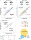
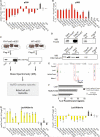
Figure 1. Foxd3 acts mostly as a transcriptional repressor in mESC
(A) RNA-seq experiments in Foxd3fl/fl;Cre-ER mESC treated with TM for three days (Foxd3−/−) or left untreated (Foxd3fl/fl). Mouse genes were plotted according to average normalized RNA-seq read counts in Foxd3−/− and Foxd3fl/fl cells. (B) RNA-seq experiments in tetON Foxd3 and WT mESC treated with Dox for three days. (C) Mouse genes considered as differentially expressed in Foxd3−/− mESC are plotted according to the RNA-seq read counts in Foxd3−/− and Foxd3fl/fl mESC and color-coded according to expression changes in tetON Foxd3 cells (purple for downregulated genes; green for upregulated genes). (D) ChIP-seq profiles generated in Foxd3-FH mESC with anti-HA and anti-Flag antibodies around a representative locus (i.e. Tfap2c). (E) Overrepresented gmotifs enriched at Foxd3-bound regions based on matches to known TF consensus binding sequences (top) or through de novo motif analysis (bottom). (F) Venn-diagram representing the overlaps between genes bound by Foxd3 and genes considered as either up or downregulated in Foxd3−/− mESC. P-values in (C) and (F) were calculated using hypergeometric tests. See also Figure S1, Data S1-2.
Figure 2
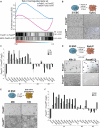
Figure 2. Foxd3 is required for the exit from naïve pluripotency
(A) GSEA for the 500 most downregulated genes during the differentiation of 2i ESC into EpiLC (Hayashi et al., 2011) with respect to the global transcriptional changes observed in Foxd3−/− Vs Foxd3fl/fl or tetON Foxd3 Vs WT mESC. ES: enrichment score. (B) Brightfield images are shown for: Foxd3fl/fl;Cre-ER cells adapted to 2i+LIF conditions (2i ESC) and then treated with TM for three days (Foxd3−/−) or left untreated (Foxd3fl/fl) (left panels); Foxd3−/− and Foxd3fl/fl cells after differentiation into EpiLC (right panels). (C) Foxd3fl/fl;Cre-ER 2i ESC were treated as in (B) and transcriptional changes between Foxd3−/ and Foxd3fl/fl cells during differentiation into EpiLC are presented in log2 scale. #1: naïve pluripotency and early germline genes repressed by Foxd3 according to RNA-seq data in Foxd3−/− mESC; #2 pluripotency genes not repressed by Foxd3 and #3: primed pluripotency genes. (D) Foxd3fl/fl;Cre-ER 2i ESC were treated with TM or left untreated, differentiated into EpiLC for three days and then re-plated under “2i+LIF”. Left panels show Foxd3−/− and Foxd3fl/fl cells after 48 hours of being re-plated in “2i+LIF”. The Right panel shows alkaline phosphatase staining of Foxd3−/− and Foxd3fl/fl cells after two passages in “2i+LIF”. (E) Foxd3fl/fl;Cre-ER 2i ESC were pre-treated with TM for 12 hours (Foxd3−/−) or left untreated (Foxd3fl/fl) and then released from 2i+LIF for 48 (left panels) or 72 hours (right panels). TM was maintained during differentiation. (F) Foxd3fl/fl;Cre-ER 2i ESC were treated as in (E) and transcriptional changes between Foxd3−/ and Foxd3fl/fl after release from “2i+LIF” are presented in log2 scale. See also Figures S2-3, Tables S1-2.
Figure 3
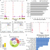
Figure 3. Foxd3 preferentially binds to active enhancers in mESC
(A) ChIP-seq profiles for the indicated proteins around a representative locus (i.e. Tfap2c). (B) Percentage of Foxd3−/− Up/EpiLC Down (Foxd3 bound regions associated with genes upregulated in Foxd3−/− mESC and downregulated during the differentiation of 2i ESC into EpiLC) and of all Foxd3 bound regions that are also bound by the indicated proteins. * statistically significant (p>0.0001) overlap. (C-D) Average ChIP-seq profiles for p300, H3K27ac and H3K4me1 in mESC around the central position of (C) all Foxd3 bound regions and (D) Foxd3−/− Up/EpiLC Down regions. (E) ChIP-seq profiles for Foxd3 in SL mESC and for p300 and H3K27ac in both 2i ESC and EpiLC around a representative locus (i.e. Tfap2c). (F) Expression levels measured as FPKMs (fragments per kilobase of exon per million fragments mapped) for all mouse genes or for genes considered as bound by Foxd3. P-value calculated using a non-paired Wilcoxon test. (G) Distances between Foxd3 bound regions and their closest ENSEMBL gene TSSs. (H) Functional annotation of Foxd3 bound regions according to GREAT analysis.
Figure 4
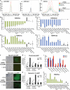
Figure 4. Foxd3 mediates the decommissioning of a subset of naïve pluripotency enhancers
(A) Average p300 and H3K27ac ChIP-seq profiles in 2i ESC and EpiLC are shown around three different subsets of TF bound regions: (i) Foxd3−/− Up/EpiLC Down; (ii) Foxd3 all (all regions bound by Foxd3 in mESC); (iii) Pou5f1 all (all regions bound by Pou5f1/Oct4 in mESC). (B-E) ChIP-qPCR analysis was performed for H3K27ac and H3K4me2 in (B-C) tetON Foxd3 and WT mESC treated with Dox for three days and (D-E) Foxd3fl/fl;Cre-ER mESC treated with TM for three days (Foxd3−/−) or left untreated (Foxd3fl/fl). ChIP signals were calculated as % of input and then normalized to the average ChIP signals obtained at two intergenic control regions (ctrl1, ctrl2). ChIP signal differences are presented in log2 scale. Foxd3 bound regions are named based on their associated genes and the distance to their TSS in Kb. Nudcd3(+19.5) and Slc25a46(−40.5) correspond to Oct4 bound genomic regions weakly bound by Foxd3 and overlapping enhancers active in both 2i ESC and EpiLC. (F) Reporter cell lines were established for two Foxd3 bound regions (Tbx3 (+9.4) and Nanos3 (−1.1)) in tetON Foxd3 mESC. GFP signals were assayed in cells that were treated with Dox (+Dox) for three days or left untreated (−Dox). (G-H) Transcriptional changes in Foxd3 and GFP levels for Tbx3(+9.4) and Nanos3(−1.1) reporter cell lines generated in tetON Foxd3 (lines #1 and #2) or WT mESC. P-values were calculated using one-tailed t-tests. (I) Changes in eRNA levels between Foxd3fl/fl;Cre-ER 2i ESC pre-treated with TM for 12 hours (Foxd3−/−) or left untreated (Foxd3fl/fl) before being released from 2i+LIF conditions for 48 hours. eRNA levels were normalized to that of two housekeeping genes (Hprt1 and Eef1a) and measured both upstream (5’, blue) and downstream (3’, red) of the indicated regions. (J) ChIP-qPCR analysis for H3K27ac in Foxd3fl/fl;Cre-ER treated like in (I). * statistically significant differences (p<0.05; fold-change (FC)>1.5 Up or Down (in log2, FC>0.58 or <-0.58)) in ChIP signals or eRNA levels between cells being compared in each case. P-values calculated using one-tailed t-test. # genomic regions considered as bound by Foxd3 according to HA ChIP-seq and just below the calling cut-off in Flag ChIP-seq (Data S2). See also Figures S4-5.
Figure 5
Figure 5. Foxd3 shifts the balance of co-activator and co-repressor proteins bound to naïve pluripotency enhancers
(A-B) ChIP-qPCR analysis for p300 in (A) tetON Foxd3 and WT mESC treated with Dox for three days and (B) Foxd3fl/fl;Cre-ER mESC treated with TM for three days (Foxd3−/−) or left untreated (Foxd3fl/fl). (C) Strategy used to identify Foxd3 interacting proteins. FT (flow-through); IP (immunoprecipitation). (D) IP with anti-Flag antibody was performed in FH-Foxd3 and WT mESC. Protein levels were detected by Western blotting using antibodies against Foxd3 or Lsd1/Kdm1a. (E) ChIP-seq profiles for Foxd3 and the indicated NuRD subunits around a representative locus (i.e. Tfap2c). (F) Percentage of all or Foxd3−/− Up/EpiLC Down Foxd3 bound regions that are also bound by the indicated NuRD subunits in mESC. * statistically significant (p<0.0001) overlap. (G-H) ChIP-qPCR analysis for Lsd1 in (G) tetON Foxd3 and WT mESC treated with Dox for three days and (H) Foxd3fl/fl;Cre-ER mESC treated with TM for three days (Foxd3−/−) or left untreated (Foxd3fl/fl). In (A-B) and (G-H) * statistically significant differences in ChIP signals as described in Figure 4. See also Figure S6, Table S3.
Figure 6
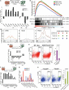
Figure 6. Silencing of Foxd3 is required for PGCLC specification
(A) PGCLC were sorted by FACS using antibodies against ITGB3 and SSEA1 (Hayashi et al., 2011). Cells displaying high signals for both ITGB3 and SSEA1 were considered as PGCLC (PGCLC +), while the remaining ones were not (PGCLC −). Transcriptional changes in PGCLC+ and PGCLC− cells relative to EpiLC are presented in log2 scale. (B) GSEA for three different set of genes (PGC core: “core” upregulated genes during PGC specification (Nakaki et al., 2013); PGCLC Up: top 500 upregulated genes during the differentiation of EpiLC into PGCLC (Hayashi et al., 2011); PGC Up: top 500 upregulated genes in E9.5 PGC compared to E5.75 epiblast (Hayashi et al., 2011)) with respect to the global transcriptional changes observed between Foxd3−/− and Foxd3fl/fl mESC. (C) Average H3K27ac ChIP-seq signal profiles in 2i ESC, EpiLC and PGCLC around three different subsets of genomic regions: (i) Foxd3−/− Up/PGCLC Up (Foxd3 bound regions associated with genes upregulated in Foxd3−/− mESC and during PGCLC differentiation); (ii) Foxd3 all; (iii) Pou5f1 all. (D) EpiLC derived from tetON Foxd3 and WT 2i ESC were treated with Dox at the beginning (day 0) and after two days (day 2) of PGCLC differentiation. After four days, the resulting cell aggregates were used to investigate the transcriptional changes between tetON Foxd3 and WT cells. (E) tetON Foxd3 and WT 2iESC were differentiated into PGCLC as described in (D). After four days, PGCLC were quantified by FACS. Representative experiments in WT and tetON Foxd3 cells and the average results from five independent quantifications are presented. P-value was calculated using paired t-test. (F) EpiLC derived from Foxd3fl/fl;Cre-ER 2i ESC were treated with TM at the beginning (day 0) and after two days (day 2) of PGCLC differentiation (Foxd3−/−) or left untreated (Foxd3fl/fl). After four days, the resulting cell aggregates were used to investigate the transcriptional changes between Foxd3−/− and Foxd3fl/fl cells. (G) Transcriptional changes in eRNA levels between PGCLC aggregates derived as described in (F). * denotes statistically significant differences in eRNA levels as described in Figure 4. (H) Foxd3fl/fl;Cre-ER 2i ESC were differentiated into PGCLC as described in (F). After four days, the number of PGCLC was quantified by FACS. Representative experiments in Foxd3fl/fl and Foxd3−/− cells and the average results from five independent quantifications are presented. P-value was calculated using paired t-test. See also Figure S7.
Figure 7
Figure 7. Proposed model for Foxd3 regulatory function during mouse peri-implantation development
Our data suggests that a wave of activation-deactivation of Foxd3 is crucial for the exit from naïve pluripotency and subsequent PGC specification. Foxd3 executes its regulatory function through enhancer decommissioning and consequent repression of relevant target genes.

Comment in

Similar articles

Cited by

References

    1. Andersson R, Gebhard C, Miguel-Escalada I, Hoof I, Bornholdt J, Boyd M, Chen Y, Zhao X, Schmidl C, Suzuki T, et al. An atlas of active enhancers across human cell types and tissues. Nature. 2014;507:455–461. - PMC - PubMed
    1. Betschinger J, Nichols J, Dietmann S, Corrin PD, Paddison PJ, Smith A. Exit from pluripotency is gated by intracellular redistribution of the bHLH transcription factor Tfe3. Cell. 2013;153:335–347. - PMC - PubMed
    1. Bonn S, Zinzen RP, Girardot C, Gustafson EH, Perez-Gonzalez A, Delhomme N, Ghavi-Helm Y, Wilczynski B, Riddell A, Furlong EEM. Tissue-specific analysis of chromatin state identifies temporal signatures of enhancer activity during embryonic development. Nat. Genet. 2012;44:148–156. - PubMed
    1. Borgel J, Guibert S, Li Y, Chiba H, Schubeler D, Sasaki H, Forne T, Weber M. Targets and dynamics of promoter DNA methylation during early mouse development. Nat. Genet. 2010;42:1093–1100. - PubMed
    1. Brackertz M, Boeke J, Zhang R, Renkawitz R. Two highly related p66 proteins comprise a new family of potent transcriptional repressors interacting with MBD2 and MBD3. J. Biol. Chem. 2002;277:40958–40966. - PubMed

Publication types

MeSH terms

Substances