iBet uBet web content aggregator. Adding the entire web to your favor.


Link to original content: https://pubmed.ncbi.nlm.nih.gov/25923211/
New Information on Tataouinea hannibalis from the Early Cretaceous of Tunisia and Implications for the Tempo and Mode of Rebbachisaurid Sauropod Evolution - PubMed Skip to main page content
U.S. flag

An official website of the United States government

Dot gov

The .gov means it’s official.
Federal government websites often end in .gov or .mil. Before sharing sensitive information, make sure you’re on a federal government site.

Https

The site is secure.
The https:// ensures that you are connecting to the official website and that any information you provide is encrypted and transmitted securely.

Access keys NCBI Homepage MyNCBI Homepage Main Content Main Navigation
. 2015 Apr 29;10(4):e0123475.
doi: 10.1371/journal.pone.0123475. eCollection 2015.

New Information on Tataouinea hannibalis from the Early Cretaceous of Tunisia and Implications for the Tempo and Mode of Rebbachisaurid Sauropod Evolution

Affiliations

New Information on Tataouinea hannibalis from the Early Cretaceous of Tunisia and Implications for the Tempo and Mode of Rebbachisaurid Sauropod Evolution

Federico Fanti et al. PLoS One. .

Abstract

The rebbachisaurid sauropod Tataouinea hannibalis represents the first articulated dinosaur skeleton from Tunisia and one of the best preserved in northern Africa. The type specimen was collected from the lower Albian, fluvio-estuarine deposits of the Ain el Guettar Formation (southern Tunisia). We present detailed analyses on the sedimentology and facies distribution at the main quarry and a revision of the vertebrate fauna associated with the skeleton. Data provide information on a complex ecosystem dominated by crocodilian and other brackish water taxa. Taphonomic interpretations indicate a multi-event, pre-burial history with a combination of rapid segregation in high sediment supply conditions and partial subaerial exposure of the carcass. After the collection in 2011 of the articulated sacrum and proximalmost caudal vertebrae, all showing a complex pattern of pneumatization, newly discovered material of the type specimen allows a detailed osteological description of Tataouinea. The sacrum, the complete and articulated caudal vertebrae 1-17, both ilia and ischia display asymmetrical pneumatization, with the left side of vertebrae and the left ischium showing a more extensive invasion by pneumatic features than their right counterparts. A pneumatic hiatus is present in caudal centra 7 to 13, whereas caudal centra 14-16 are pneumatised by shallow fossae. Bayesian inference analyses integrating morphological, stratigraphic and paleogeographic data support a flagellicaudatan-rebbachisaurid divergence at about 163 Ma and a South American ancestral range for rebbachisaurids. Results presented here suggest an exclusively South American Limaysaurinae and a more widely distributed Rebbachisaurinae lineage, the latter including the South American taxon Katepensaurus and a clade including African and European taxa, with Tataouinea as sister taxon of Rebbachisaurus. This scenario would indicate that South America was not affected by the end-Jurassic extinction of diplodocoids, and was most likely the centre of the rapid radiation of rebbachisaurids to Africa and Europe between 135 and 130 Ma.


Conflict of interest statement

Competing Interests: The authors have declared that no competing interests exist.

Figures

Fig 1
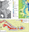
Fig 1. The Tataouine basin in southern Tunisia.
A, reference map of the Tataouine region in southern Tunisia; B, simplified geological map of the study area showing the distribution of Mesozoic deposits and the El Mra locality near the village of Bir Amir. C, detailed topographic and geological map of the El Mra locality.
Fig 2
Fig 2. Stratigraphy at the El Mra locality.
A, panoramic view of the El Mra mesa-like morphology. 1. Chenini Mbr., 2. Oum Ed Diab Mbr., 3. Keker Mbr., 4. Gattar Mbr. B, simplified field-log of the El Mra section showing the stratigraphic occurrence of vertebrate and plant remains. C, facies distribution in the Chenini-Oum Ed Diab transition. F1, high-energy, fluvial sandy bar deposits; F2, low-angle shoreface deposits; F3, fine-grained tidal deposits with flaser-like structures. D, E, F, field photographs of the different deposits exposed at the El Mra locality. T. hannibalis was recovered from the F1 beds.
Fig 3
Fig 3. Preserved elements of Tataouinea hannibalis (ONM DT 1–48).
A, quarry map showing the orientation of collected elements. B, field photograph of the elements collected at the end of 2011, and C, of elements collected in 2013. Ca, caudal vertebra 1–17; S, sacral vertebra 1–5; r il, right ilium; l il, left ilium.
Fig 4
Fig 4. Partial sacrum of Tataouinea hannibalis.
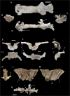
Partial sacral centra 1–4 in ventral (A), cranial (B), caudal (C) and lef lateral (D) views. Partial sacral centrum 5 in caudal (E), cranial (F), right lateral (G), ventral (H), dorsal (I) and left lateral (J) views. Scale bars: 10 cm. Abbreviations: emp, extramural pneumatisation; fo, fossa; iss, interspinal suture scar; ll, lateral lamina; sr, sacral ribs.
Fig 5
Fig 5. Sacral neural spines 4 and 5 of Tataouinea hannibalis.
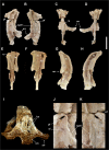
Sacral neural spine 4 in left lateral (A), right lateral (B), cranial (C) and caudal (D) views. Sacral neural spine 5 in caudal (E), cranial (F), right lateral (G) and left lateral (H) views. Cross section of sacral neural spine 4 (I). Details of sacral neural spine 4 pneumatisation in left lateral (J) and right lateral (K) views. Scale bar: 10 cm. Abbreviation: arr, alar ramus of rib; ca, camera; cm, camellae; fo, fossa; ics, intercamerate septum; pf, pneumatic foramen; ru, rugosities; sf, semilunate fossa; spdl, spinodiapophyseal lamina.
Fig 6
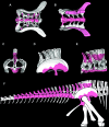
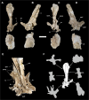
Fig 6. Sacrum with associated ilia, and reconstruction of known elements of Tataouinea hannibalis.
Sacrum in dorsal view (A), ventral view (B), caudal view (C), right lateral view with ilium associated (D) and with ilium removed (E). In (B), the peduncles of the right ilium are removed. Skeletal reconstruction of caudosacral region of Tataouinea hannibalis (F). Recovered elements in colour. A full skeletal reconstruction is shown in S1 File.
Fig 7
Fig 7. Partial proximal caudal vertebrae of Tataouinea hannibalis.
Partial caudal neural spine 1 in right lateral view (A). Partial caudal centrum 1 in right lateral view (B). Partial neural arch 3 in right lateral (C) and proximal (D) views. Partial caudal neural arch 4 in right lateral view (E). Scale bars: A-D: 10 cm, E: 10 cm. Abbreviations: fo, fossa; ll, lateral lamina; pl, pleurocoel; prsl, prespinal lamina; spdl, spinodiapophyseal lamina; spol, spinopostzygapophyseal lamina; sprl, spinoprezygapophyseal lamina.
Fig 8
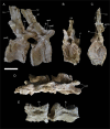
Fig 8. Caudal vertebra 5 of Tataouinea hannibalis.
Vertebra in proximal (A), left lateral (B), distal (C), right lateral (D) views. Detail of neural arch in right proximodorsal view (D). Cross section shapes of neural arch (shown in proximal view) in six points indicated by arrows (proximal is bottom). Scale bar: 10 cm. Abbreviations: fo, fossa; hr, hyposphenal ridge; ll, lateral lamina; nc, neural canal; pl, pleurocoel; pozp, postzygapophysis pathology; psl, prespinal lamina; pz, prezygapophysis; pzdl, prezygodiapophyseal lamina; ri, rib; spol, spinopostzygapophyseal lamina; sprl, spinoprezygapophyseal lamina.
Fig 9
Fig 9. Caudal vertebra 6 of Tataouinea hannibalis.
Vertebra in left lateral (A) and distal (B) views. Scale bar: 10 cm. Abbreviations: hr, hyposphenal ridge; nc, neural canal; podl, postzygodiapophyseal lamina; ri, rib; spof, spinopostzygapophyseal fossa; spol, spinopostzygapophyseal lamina.
Fig 10
Fig 10. Caudal vertebra 7 of Tataouinea hannibalis.
Vertebra in right lateral (A), proximal (B) and distal (C) views. Scale bar: 10 cm. Abbreviations: col, collapsed area; fo, fossa; hr, hyposphenal ridge; nc, neural canal; pcdl, posterior centrodiapophyseal lamina; podl, postzygodiapophyseal lamina; pz, prezygapophysis; pzdl, prezygodiapophyseal lamina; ri, rib; spof, spinopostzygapophyseal fossa; sprl, spinoprezygapophyseal lamina.
Fig 11
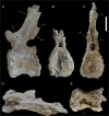
Fig 11. Caudal vertebrae 8 and 9 of Tataouinea hannibalis.
Vertebrae in right lateral (A), proximal (B), distal (C), dorsal (D) and ventral (E) views. Scale bar: 10 cm. Abbreviations: al, accessory laminae; fo, fossa; hr, hyposphenal ridge; ns, neural spine; pcdl, posterior centrodiapophyseal lamina; pz, prezygapophysis; pz10, fragment of caudal 10 right prezygapophysis; pzdl, prezygodiapophyseal lamina; ri, ribs; ru, interspinal rugosity; sprl, spinoprezygapophyseal lamina.
Fig 12
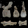
Fig 12. Caudal vertebra 10 of Tataouinea hannibalis.
Vertebra in left lateral (A), proximal (B), distal (C), dorsal (D) and ventral (E) views. Scale bar: 10 cm. Abbreviations: fo, fossa; hr, hyposphenal ridge; ipf, interprezygapophyseal fossa; nc, neural canal; poz, postzygapophysis; pz11, fragment of caudal 11 left prezygapophysis; ru, rugosities.
Fig 13
Fig 13. Caudal vertebra 11 of Tataouinea hannibalis.
Vertebra in left lateral (A), proximal (B), distal (C), dorsal (D) and ventral (E) views. Scale bar: 10 cm. Abbreviation: ca, camerae; fo, fossa; ipf, interprezygapophyseal fossa; nc, neural canal; poz, postzygapophysis; poz10, fragment of caudal 10 right postzygapophysis; ru, rugosities.
Fig 14
Fig 14. Caudal vertebra 12 of Tataouinea hannibalis.
Vertebra in right lateral (A), proximal (B), distal (C), dorsal (D) and ventral (E) views. Scale bar: 10 cm. Abbreviation: fo, fossa; hr, hyposphenal ridge; nc, neural canal; ns, neural spine; poz, postzygapophysis; poz11, fragment of caudal 11 postzygapophysis; pz, prezygapophysis; ru, rugosities; sprf, spinoprezygapophyseal fossa.
Fig 15
Fig 15. Caudal vertebrae 13 and 14 of Tataouinea hannibalis.
Vertebrae in right lateral (A), proximal (B), distal (C), dorsal (D) and ventral (E) views. Scale bar: 10 cm. Abbreviations: fo, fossa; hr, hyposphenal ridge; ns, neural spine; poz, postzygapophysis; pz, prezygapophysis; ru, rugosities; sprf, spinoprezygapophyseal fossa.
Fig 16
Fig 16. Caudal vertebra 15 of Tataouionea hannibalis.
Vertebra in right lateral (A), proximal (B), distal (C), dorsal (D) and ventral (E) views. Scale bar: 10 cm. Abbreviations: col, collapsed area; fo, fossa; hr, hyposphenal ridge; ns, neural spine; poz, postzygapophysis; poz14, right caudal 14 postzygapophysis fragment; sprf, spinoprezygapophyseal fossa;.
Fig 17
Fig 17. Caudal vertebra 16 of Tataouinea hannibalis.
Vertebra in right lateral (A), proximal (B), distal (C), dorsal (D) and ventral (E) views. Scale bar: 10 cm. Abbreviations: ca, camerae; col, collapsed area; fo, fossae; hr, hyposphenal ridge; nc, neural canal; ns, neural spine; poz, postzygapophysis; pz, prezygapophysis; ru, rugosities.
Fig 18
Fig 18. Caudal vertebra 17 of Tataouinea hannibalis.
Vertebra in left lateral (A) and distal (B) views. Scale bar: 10 cm. Abbreviations: ns, neural spine; poz, postzygapophysis; pz, prezygapophysis.
Fig 19
Fig 19. Pelvic elements of Tataouinea hannibalis.
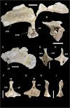
Left ilium in lateral view (A). Left pubic peduncle in distal/ventral view (B). Right preacetabular process of ilium in lateral view (C). Right ischium in medial (D) and lateral (E) views. Left ischium in lateral (F) and medial (G) views. Right ischium in proximal (acetabular) view (H). Left ischium in proximal (acetabular) view (I). Scale bar: 10 cm. Abbreviations: am, acetabular margin; col, collapsed area; ic, internal chamber; ilf, iliac facet; pf, pneumatic foramen; prp, preacetabular process; pup, pubic peduncle; se, septa.
Fig 20
Fig 20. Left ischium of Tataouinea hannibalis.
Ischium in lateral (A), medial (B), cranial (C), caudal (D), proximal/dorsal (E), distal/ventral (F) views. Scale bar: 10 cm. Abbreviations: am, acetabular margin; ilf, iliac facet; mf, medial fossa; ms, muscle scar; pf, pneumatic foramen; puf, pubic facet.
Fig 21
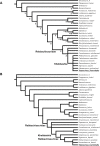
Fig 21. Phylogenetic relationships among rebbachisaurids.
Strict consensus topology under equal weighting (A) and under implied weighting (B) of the shortest trees recovered by the parsimony analyses of the dataset. Numbers adjacent to nodes in the equally weighted analysis tree indicate Decay Index values >1.
Fig 22
Fig 22. BBM palaeogeographyc analysis of Rebbachisauridae.
Time-calibrated palaeobiogeography of eusauropods focusing on rebbachisaurids (above) and result of the time-event algorithm test on Rebbachisauridae (below), based on the BBM analysis of the MCCT recovered by Bayesian inference. Values at nodes indicate posterior probability values >0.5. Abbreviations: A, Asia; B, Europe; C, North America; D, Africa; E, South America. Black circles indicate uncertain optimization.
Fig 23
Fig 23. S-DIVA palaeogeographyc analysis of Rebbachisauridae.
Time-calibrated palaeobiogeography of eusauropods focusing on rebbachisaurids (above) and result of the time-event algorithm test on Rebbachisauridae (below), based on the S-DIVA analysis of the MCCT recovered by Bayesian inference. Values at nodes indicate posterior probability values >0.5. Abbreviations: A, Asia; B, Europe; C, North America; D, Africa; E, South America. Black circles indicate uncertain optimization.

Similar articles

Cited by

References

    1. Fanti F, Cau A, Hassine M, Contessi M. A new sauropod dinosaur from the Early Cretaceous of Tunisia with extreme avian-like pneumatization. Nature Communications 2013;4:2028, 7 pp. 10.1038/ncomms3028 - DOI - PubMed
    1. Pervinquière L. Sur la géologie de l’extrême Sud tunisien et de la Tripolitaine. Bulletin de la Société Géologique de France XII 1912; (4), 160.
    1. Kilian C. Des principaux complexes continentaux du Sahara. Comptes Rendus de la Société Géologique de France 1931;9: 109–111.
    1. Kilian C. Esquisse géologique du Sahara français à l’est du 6ème degré de longitude, Chronique des Mines et de la Colonisation 1937; 58, 21–22 Map I/4.000.000 Paris.
    1. de Lapparent AF. Découverte de Dinosauriens, associes a une faune de Reptiles et de Poissons, dans le Crétacé inferieur de l”extrême Sud tunisien. Comptes Rendus Académie des Sciences Paris 1951;232: 1430–1432.

Publication types

Grants and funding

This research was conducted in collaboration with the Office National Des Mines, Tunis, and was financially supported by Fondazione Alma Mater (Professor G. Gabbianelli), the Integrated Geoscience Research Group (Department of Earth and Geo-Environmental Sciences, University of Bologna, Italy), the TITAN Project (Professor L. Cantelli) and the Museo Geologico Giovanni Capellini (Bologna, Italy) who supported field and laboratory activities. The funders had no role in study design, data collection and analysis, decision to publish, or preparation of the manuscript.

LinkOut - more resources