iBet uBet web content aggregator. Adding the entire web to your favor.


Link to original content: https://pubmed.ncbi.nlm.nih.gov/25855243
Incorporation of spike and membrane glycoproteins into coronavirus virions - PubMed Skip to main page content
U.S. flag

An official website of the United States government

Dot gov

The .gov means it’s official.
Federal government websites often end in .gov or .mil. Before sharing sensitive information, make sure you’re on a federal government site.

Https

The site is secure.
The https:// ensures that you are connecting to the official website and that any information you provide is encrypted and transmitted securely.

Access keys NCBI Homepage MyNCBI Homepage Main Content Main Navigation
Review
. 2015 Apr 3;7(4):1700-25.
doi: 10.3390/v7041700.

Incorporation of spike and membrane glycoproteins into coronavirus virions

Affiliations
Review

Incorporation of spike and membrane glycoproteins into coronavirus virions

Makoto Ujike et al. Viruses. .

Abstract

The envelopes of coronaviruses (CoVs) contain primarily three proteins; the two major glycoproteins spike (S) and membrane (M), and envelope (E), a non-glycosylated protein. Unlike other enveloped viruses, CoVs bud and assemble at the endoplasmic reticulum (ER)-Golgi intermediate compartment (ERGIC). For efficient virion assembly, these proteins must be targeted to the budding site and to interact with each other or the ribonucleoprotein. Thus, the efficient incorporation of viral envelope proteins into CoV virions depends on protein trafficking and protein-protein interactions near the ERGIC. The goal of this review is to summarize recent findings on the mechanism of incorporation of the M and S glycoproteins into the CoV virion, focusing on protein trafficking and protein-protein interactions.


Figures
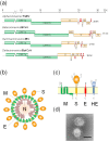
Figure 1
Figure 1
(a) Genome organizations of various Coronavirus genera. The first two-thirds of the genome contains open reading frame (ORF)1a and ORF1b, which encode the replicase/transcriptase proteins (Green). The remaining one-third of the genome encodes four structural proteins: spike (S), envelope (E), membrane (M), and nucleocapsid (N) proteins (Orange). Some betaoronaviruses have an additional hemagglutinin-esterase (HE) gene (Orange). The genome of each genus or species has a set of unique accessory proteins (red); (b) Schematic diagrams of coronavirus virions; (c) The topology of the four structural envelope proteins. All proteins are depicted as monomers, but the S and HE proteins form homotrimers and homodimers, respectively. Oligosaccharides are shown on the M, S, and HE proteins. Although a number are omitted, the S and HE proteins contain 21 to 35 and 9 (BCoV HE) potential N-glycosylation sites, respectively; (d) Electron micrograph of SCoV. Bar: 100 nm. (EM image courtesy of Dr. Nagata at National Institute of Infectious Diseases).
Figure 2
Figure 2
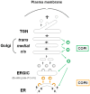
Cisternal maturation and stable (ER)-Golgi intermediate compartment (ERGIC) model. The protein in COPII vesicles (Orange) buds from ER-exit sites to the ERGIC, which is a stable compartment in mammalian cells, and subsequently to the cis-Golgi via a second undefined vesicular transport system (Grey) [53,61]. These vesicles fuse homotypically to construct new cis-cisternae, which are not stable compartments. They move and mature from cis to trans, and then breakdown into transport carriers at the TGN. Transport between the ER and Golgi is controlled by COPI vesicles (Green) in retrograde, and COPIl vesicles in anterograde, transport [55,56,58]. (Figure is modified, based on figure of Glick et al., reference 56).
Figure 3
Figure 3
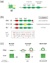
(a) Topology and schematic diagram of coronaviruses (CoV) M proteins. Three TM domains were assigned to tm1, tm2, and tm3 regions; (b) Trafficking signals of CoV-M proteins. Red box shows the identified intracellular retention signal, and the yellow box the plasma membrane targeting signal. The tm1 regions of SCoV- and infectious bronchitis virus (IBV), but not MHV-, M proteins are sufficient for intracellular retention (Top). Amino acid sequences of the tm1 regions of three CoV-M protein (Bottom). Conserved amino acids are shown in red. Asterisks indicate the uncharged polar residues critical for intracellular retention of VSV-G with tm1 regions of IBV-M [82]; (c) Minimum requirement for virus-like particle (VLP) formation. Coexpression of M and E proteins, but not M protein alone, resulted in formation of VLPs. E proteins might cause membrane bending or scission at the budding site. N protein likely assists VLP formation. In contrast, SCoV-M protein alone resulted in production of VLP, albeit at a low density. The minimum requirement for SCoV VLP assembly is controversial; (d) The importance of M-M interactions via multiple contact sites in the overall domain for VLP incorporation. Assembly-incompetent MHV-M proteins (light blue) lacking parts of the TM domain, the amphipathic domain, or the C-terminal domain, or M proteins in which the N-terminal domain has been replaced by exogenous proteins, lack VLP formation ability. An increase in the quantity of assembly-competent M proteins (green) promotes VLP formation and provides more opportunities for the binding and capture of incompetent M proteins into VLPs, resulting in an increase in the co-incorporation of incompetent M proteins into VLPs or virions [46]. E proteins are omitted; (e,f) Lattice-like matrix model of M protein in edge (e) and axial (f) views. M protein would form a lattice-like matrix within the envelope. RNPs would interact with M proteins to be packaged into virions, and S proteins are accommodated within spaces of this lattice via interaction with M proteins. Small numbers of E proteins are inserted into the other spaces within the lattice.
Figure 3
Figure 3
(a) Topology and schematic diagram of coronaviruses (CoV) M proteins. Three TM domains were assigned to tm1, tm2, and tm3 regions; (b) Trafficking signals of CoV-M proteins. Red box shows the identified intracellular retention signal, and the yellow box the plasma membrane targeting signal. The tm1 regions of SCoV- and infectious bronchitis virus (IBV), but not MHV-, M proteins are sufficient for intracellular retention (Top). Amino acid sequences of the tm1 regions of three CoV-M protein (Bottom). Conserved amino acids are shown in red. Asterisks indicate the uncharged polar residues critical for intracellular retention of VSV-G with tm1 regions of IBV-M [82]; (c) Minimum requirement for virus-like particle (VLP) formation. Coexpression of M and E proteins, but not M protein alone, resulted in formation of VLPs. E proteins might cause membrane bending or scission at the budding site. N protein likely assists VLP formation. In contrast, SCoV-M protein alone resulted in production of VLP, albeit at a low density. The minimum requirement for SCoV VLP assembly is controversial; (d) The importance of M-M interactions via multiple contact sites in the overall domain for VLP incorporation. Assembly-incompetent MHV-M proteins (light blue) lacking parts of the TM domain, the amphipathic domain, or the C-terminal domain, or M proteins in which the N-terminal domain has been replaced by exogenous proteins, lack VLP formation ability. An increase in the quantity of assembly-competent M proteins (green) promotes VLP formation and provides more opportunities for the binding and capture of incompetent M proteins into VLPs, resulting in an increase in the co-incorporation of incompetent M proteins into VLPs or virions [46]. E proteins are omitted; (e,f) Lattice-like matrix model of M protein in edge (e) and axial (f) views. M protein would form a lattice-like matrix within the envelope. RNPs would interact with M proteins to be packaged into virions, and S proteins are accommodated within spaces of this lattice via interaction with M proteins. Small numbers of E proteins are inserted into the other spaces within the lattice.
Figure 4
Figure 4
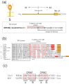
(a) Schematic diagram of CoV-S proteins and CT domain amino acid sequences. The CoV S2 domain can be further divided into three subdomains; a large ectodomain, a single TM domain, and a CT domain (top). The deduced CT domain sequence of MHV-S (A59) protein comprises two subdomains: a cysteine-rich motif (CRM) and charge-rich domains, which partially overlap (bottom); (b) C-terminal ends of 10 CoV-S proteins. Amino acid sequence alignment was performed by CLUSTALW. Shaded box indicates the deduced TM domain. Cysteine residues in CRM are shown in red. Orange and red boxes indicate potential ER retrieval signals (KxHxx- or KKxx-motif) and tyrosine-dependent localization signals/internalization signals (YxxФ motif, where Ф can be F, I, L, M or V), respectively. (c) Comparison of the CRM domains of SCoV- and MHV-S. The CRM can be further divided into four subclusters.

Similar articles

Cited by

References

    1. Hamre D., Procknow J.J. A new virus isolated from the human respiratory tract. Proc. Soc. Exp. Biol. Med. 1966;121:190–193. doi: 10.3181/00379727-121-30734. - DOI - PubMed
    1. McIntosh K., Dees J.H., Becker W.B., Kapikian A.Z., Chanock R.M. Recovery in tracheal organ cultures of novel viruses from patients with respiratory disease. Proc. Natl. Acad. Sci. USA. 1967;57:933–940. doi: 10.1073/pnas.57.4.933. - DOI - PMC - PubMed
    1. Tyrrell D.A., Bynoe M.L. Cultivation of a novel type of common-cold virus in organ cultures. Br. Med. J. 1965;1:1467–1470. doi: 10.1136/bmj.1.5448.1467. - DOI - PMC - PubMed
    1. van der Hoek L., Pyrc K., Jebbink M.F., Vermeulen-Oost W., Berkhout R.J., Wolthers K.C., Wertheim-van Dillen P.M., Kaandorp J., Spaargaren J., Berkhout B. Identification of a new human coronavirus. Nat. Med. 2004;10:368–373. - PMC - PubMed
    1. Woo P.C., Lau S.K., Tsoi H.W., Huang Y., Poon R.W., Chu C.M., Lee R.A., Luk W.K., Wong G.K., Wong B.H., et al. Clinical and molecular epidemiological features of coronavirus hku1-associated community-acquired pneumonia. J. Infect. Dis. 2005;192:1898–1907. doi: 10.1086/497151. - DOI - PMC - PubMed

Publication types

Substances