iBet uBet web content aggregator. Adding the entire web to your favor.


Link to original content: https://pubmed.ncbi.nlm.nih.gov/25662009
Chromothriptic cure of WHIM syndrome - PubMed Skip to main page content
U.S. flag

An official website of the United States government

Dot gov

The .gov means it’s official.
Federal government websites often end in .gov or .mil. Before sharing sensitive information, make sure you’re on a federal government site.

Https

The site is secure.
The https:// ensures that you are connecting to the official website and that any information you provide is encrypted and transmitted securely.

Access keys NCBI Homepage MyNCBI Homepage Main Content Main Navigation
Case Reports
. 2015 Feb 12;160(4):686-699.
doi: 10.1016/j.cell.2015.01.014. Epub 2015 Feb 5.

Chromothriptic cure of WHIM syndrome

Affiliations
Case Reports

Chromothriptic cure of WHIM syndrome

David H McDermott et al. Cell. .

Abstract

Chromothripsis is a catastrophic cellular event recently described in cancer in which chromosomes undergo massive deletion and rearrangement. Here, we report a case in which chromothripsis spontaneously cured a patient with WHIM syndrome, an autosomal dominant combined immunodeficiency disease caused by gain-of-function mutation of the chemokine receptor CXCR4. In this patient, deletion of the disease allele, CXCR4(R334X), as well as 163 other genes from one copy of chromosome 2 occurred in a hematopoietic stem cell (HSC) that repopulated the myeloid but not the lymphoid lineage. In competitive mouse bone marrow (BM) transplantation experiments, Cxcr4 haploinsufficiency was sufficient to confer a strong long-term engraftment advantage of donor BM over BM from either wild-type or WHIM syndrome model mice, suggesting a potential mechanism for the patient's cure. Our findings suggest that partial inactivation of CXCR4 may have general utility as a strategy to promote HSC engraftment in transplantation.


Conflict of interest statement

Conflict of Interest

A provisional patent on CXCR4 knock down as a method to enhance HSC engraftment has been filed by the US government with DHM, QL, MS, JG, HLM, and PMM as inventors. The authors confirm that there are no other conflicts of interest.

Figures
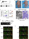
Figure 1
Figure 1. Long-term clinical remission of WHIM syndrome and evidence for somatic mosaicism in patient WHIM-09
(A) Family Pedigree. Squares, males; circles, females; shaded symbols, WHIM syndrome; arrow, index patient WHIM-09. (B) Spontaneous and complete remission of warts in patient WHIM-09. According to patient WHIM-09, through her fourth decade of life she had had extensive warts on her hands, similar to her daughters, (illustrated here for 24 year old daughter WHIM-11) that spontaneously resolved. (C) Spontaneous sustained correction of neutropenia and monocytopenia in patient WHIM-09. WBC, white blood cell count; ANC, absolute neutrophil count; AMC, absolute monocyte count; ALC, absolute lymphocyte count. Arrows indicate age at splenectomy; horizontal lines indicate normal range for each cell type. Note, × axis is discontinuous to show pre/post splenectomy results more clearly. (D) Normalization of bone marrow pathology in patient WHIM-09. A representative high magnification (500×) Wright-Giemsa stain of the bone marrow aspirate is shown for the index patient WHIM-09 in 1963 (Zuelzer, 1964) (reproduced with permission) and in 2013 at ages 9 and 59, respectively. Arrows in left image, eyeglass nuclei in neutrophils. (E) Polymerase chain reaction-BstUI restriction fragment length polymorphism (BstUI) analysis of genomic DNA. HD, healthy donor; WHIM-09, index patient; Cheek, cheek swab cells; PBMC, peripheral blood mononuclear cells; Fb, fibroblast; LCL, lymphoblastoid cell line; WB, whole blood; WT, wild-type allele; WHIM, CXCR4R334X allele that causes WHIM syndrome. (F) Sanger DNA sequencing analysis of whole blood DNA for affected family members in the region near nucleotide position 1000 (vertical line), the site of WHIM mutation CXCR4R334X (1000 C→T). Blue, C; Red, T; Fb, fibroblast; PMN, polymorphonuclear leukocyte (PMN). (G) Sanger DNA sequencing analysis of DNA from purified peripheral blood neutrophils, cells obtained from a buccal swab (cheek) and cultured skin fibroblasts for patient WHIM-09 in the same region as (F).
Figure 2
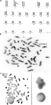
Figure 2. Massive deletion and rearrangement of one copy of chromosome 2, the location of CXCR4, in patient WHIM-09
Cytogenetics and Fluorescence in-situ hybridization (FISH) analyses were performed on cultured bone marrow cells. (A) Cytogenetics. Karyogram showing a short acrocentric chromosome 2 (arrow) with abnormal banding pattern suggesting deletions and inversions observed in all 20 metaphase cells analyzed from bone marrow. (B) FISH with intact ALK break apart probe (Abbott Molecular) signal at 2p23 on normal chromosome 2 and on long arm of abnormal chromosome 2 (der 2). (C) FISH with NMYC and CEP 2 probe set (Abbott Molecular). Left panel, Metaphase cell showing the normal chromosome 2 with intact centromere signal (red) and NMYC signal (green) at 2p24. The abnormal chromosome 2 has a portion of its centromere inverted into the long arm splitting the red signal; the green NMYC signal is absent. Right panel, Polymorphonuclear and round interphase nuclei showing the abnormal hybridization pattern with one green NMYC signal and three red CEP 2 signals. One of the red signals is smaller than the other two and is close to one of them.
Figure 3
Figure 3. Chromothripsis as the mechanism for loss of CXCR4R334X in patient WHIM-09
Purified neutrophil and cultured skin fibroblast DNA from patient WHIM-09 was isolated and subjected to whole genome sequencing with paired end analysis. (A) Linear non-proportional plot of the abnormal copy of chromosome 2 in patient WHIM-09 labeled from the p arm telomere (0) to the q arm telomere (~240) in megabases with the 18 remaining pieces arranged in their numeric order (top). Connections between these pieces are depicted by the curved lines. Note that some connections were poorly defined because of the involvement of repetitive centromeric sequence (black lines). The order and orientation of the 18 remaining pieces in the derivative chromosome are indicated at the bottom. (B) Circos plot of chromosome 2 and its normal Giemsa cytogenetic banding pattern labeled from the p arm telomere (0) to the q arm telomere (~240) in megabases. Large pieces of chromosome 2 were missing from patient WHIM-09 neutrophil DNA and the 18 remaining pieces were arranged in random order. Connections between these pieces and their orientation (inset) are depicted by the colored lines (See Figure S2 and Tables S1 and S2 for additional details). Note that 2 connections were poorly defined because of the involvement of repetitive centromeric sequence. The inner circular trace is the copy number variation data derived from microarray analysis (Figure S2A and S2B). Note that the sites of connections derived from the paired end sequencing analysis closely match the sites where copy number variation abruptly falls from 2 to 1. The location of CXCR4 is indicated at lower left. (C) Derivative chromothriptic chromosome 2. Ideogram of intact chromosome 2 (left) and a model of the Giemsa cytogenetic banding pattern and the order and orientation of the 18 pieces with the deletions called by microarray shaded in yellow (right) are shown. The resultant remaining chromosome 2 banding pattern predicted by whole genome sequencing closely matches that seen by cytogenetic analysis (see Figure 3). Note the location of CXCR4 at 2q22.1 in one of the deleted segments.
Figure 4
Figure 4. Chromothriptic CXCR4-haploinsufficient HSC replacement of the myeloid lineage, but not the lymphoid lineage, is associated with clinical remission in patient WHIM-09
(A–E) Representative results from a BstUI PCR-restriction fragment length polymorphism assay (BstUI), designed to distinguish the wild type CXCR4 allele (WT) from the CXCR4R334X WHIM allele (WHIM), as well as from a PCR assay specific for the chromothriptic chromosome (13–16 Jxn). PCR was performed on DNA obtained from the indicated donor leukocyte subsets purified either from blood using magnetic bead purification (B, E) or from a bone marrow aspirate using flow cytometric sorting (C, D). DNA was also prepared from archived WHIM-09 spleen and compared with peripheral blood PMN DNA (A), as well as from Burst-forming Unit-Erythroid colonies and compared with blood leukocyte subsets (E). WHIM-09, index patient; HD, healthy donor; Spl, spleen; PMN, polymorphonuclear leukocytes; 13–16 Jxn, PCR product specific for the chromothriptic junction between segments 13 and 16 of the chromothriptic chromosome of patient WHIM-09; PBMC, peripheral blood mononuclear cells; CD4, purified CD4+ T cells; CD8, purified CD8+ T cells; CD19 purified CD19+ B cells; CD56, purified CD56+ natural killer cells; CD14, purified CD14+ monocytes; CD34, purified CD34+ hematopoietic cells; CD3, purified CD3+ T cells; CD15, purified CD15+ neutrophils; CD16, purified CD16+ neutrophils; HSC, hematopoietic stem cells; CLP, common lymphoid precursor; CMP, common myeloid precursor; GMP, granulocyte/monocyte precursor MEP, megakaryocyte-erythroid precursor; BFU-E, Burst-forming Unit-Erythroid; CXCR4, CXCR4 amplicon not digested with BstUI. (F) Summary of myeloid/lymphoid mosaicism for CXCR4R334X in patient WHIM-09. The immunophenotype used to purify each cell type from enriched CD34+CD45+ cells is summarized next to each cell type shown. Red, positive for CXCR4R334X; green, negative for CXCR4R334X; asterisks, purified cell types directly analyzed by PCR-RFLP for the WHIM mutation.
Figure 5
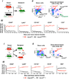
Figure 5. Cxcr4 haploinsufficiency enhances HSC engraftment during mouse bone marrow transplantation
Two types of competitive bone marrow transplantation experiments were performed: (A) Cxcr4+/o vs. Cxcr4+/S338X (mouse model of WHIM syndrome); and (B) Cxcr4+/o vs. Cxcr4+/+. For panels A and B: i) Experimental design; ii) Representative flow cytometry plots demonstrating the relative contributions of CD45 congenic markers in mixed donor bone marrow prior to transplantation (left panel) and in blood after bone marrow transplantation (right panel) in a single mouse; iii) Cell frequency data for the leukocyte subsets indicated at the top of each panel, presented as the mean ± SEM percentage (%) of total donor-derived cells for each subset (n=10 mice per data point). SEM was < 5% of the mean in all cases, and therefore is not visible for most data points. Results were verified in one and two additional independent experiments for panels A and B, respectively.
Figure 6
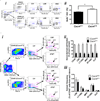
Figure 6. Cxcr4 haploinsufficiency enhances HSC proliferation and engraftment in a mouse bone marrow transplantation model
(A) Proliferation. Donor bone marrow cells with a Cxcr4+/o genotype on a homozygous CD45.2 background were mixed with donor bone marrow cells with a Cxcr4+/+ genotype on a heterozygous CD45.1/CD45.2 background (47:53) and injected intravenously into a lethally irradiated CD45.1 recipient mouse. Six days after bone marrow transplantation, each mouse was given 1.25 mg of BrdU I.P. Twenty hours later, the mice were euthanized for HSC proliferation analysis. i) Gating scheme for BrdU+ HSCs. Bone marrow cells were first gated with CD45.2 (Cxcr4+/o) and CD45.1/CD45.2 (Cxcr4+/+), then HSCs were gated as Flt3 Lin Sca1+ c-Kit+ (Flt3LSK), which includes long-term and short-term HSCs, and BrdU+ cells were quantitated. ii) Percentage of BrdU+ HSCs in each donor. Data are expressed as mean ± SEM from four mice. The experiment was repeated once with similar results. (B) Long term engraftment and differentiation. Bone marrow cells from donor mice with a Cxcr4+/o genotype on a CD45.2 background were mixed with bone marrow cells from donors with a Cxcr4+/+ genotype on a CD45.1 background (42:58) and the mixed cells were injected intravenously into lethally irradiated recipient mice. 303 days later bone marrow was harvested. i) Gating scheme for long term HSC (LT-HSC: CD34Flt3 Lin Sca1+ c-Kit+), short term HSC (ST-HSC: CD34+Flt3 Lin Sca1+ c-Kit+), multipotent progenitors (MPP: CD34+Flt3+ Lin Sca1+ c-Kit+), and common lymphoid progenitors (CLP: IL7ra+ Lin Sca1low c-Kitlow). ii) Long-term engraftment. iii) Differentiation. The distribution frequency of bone marrow cell subsets is similar for Cxcr4+/o compared to Cxcr4+/+ donor-derived cell populations. Each data point represents 10 mice presented as the mean ± SEM.

Similar articles

Cited by

References

    1. Al Ustwani O, Kurzrock R, Wetzler M. Genetics on a WHIM. British journal of haematology. 2014;164:15–23. - PMC - PubMed
    1. Bachelerie F, Ben-Baruch A, Burkhardt AM, Combadiere C, Farber JM, Graham GJ, Horuk R, Sparre-Ulrich AH, Locati M, Luster AD, et al. International Union of Pharmacology. LXXXIX. Update on the extended family of chemokine receptors and introducing a new nomenclature for atypical chemokine receptors. Pharmacological reviews. 2014;66:1–79. - PMC - PubMed
    1. Balabanian K, Brotin E, Biajoux V, Bouchet-Delbos L, Lainey E, Fenneteau O, Bonnet D, Fiette L, Emilie D, Bachelerie F. Proper desensitization of CXCR4 is required for lymphocyte development and peripheral compartmentalization in mice. Blood. 2012;119:5722–5730. - PubMed
    1. Beaussant Cohen S, Fenneteau O, Plouvier E, Rohrlich PS, Daltroff G, Plantier I, Dupuy A, Kerob D, Beaupain B, Bordigoni P, et al. Description and outcome of a cohort of 8 patients with WHIM syndrome from the French Severe Chronic Neutropenia Registry. Orphanet journal of rare diseases. 2012;7:71. - PMC - PubMed
    1. Bleul CC, Fuhlbrigge RC, Casasnovas JM, Aiuti A, Springer TA. A highly efficacious lymphocyte chemoattractant, stromal cell-derived factor 1 (SDF-1) The Journal of experimental medicine. 1996;184:1101–1109. - PMC - PubMed

Publication types

Supplementary concepts

Associated data