Subsite awareness in neuropathology evaluation of National Toxicology Program (NTP) studies: a review of select neuroanatomical structures with their functional significance in rodents
- PMID: 24135464
- PMCID: PMC3965620
- DOI: 10.1177/0192623313501893
Subsite awareness in neuropathology evaluation of National Toxicology Program (NTP) studies: a review of select neuroanatomical structures with their functional significance in rodents
Abstract
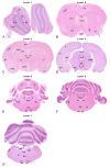
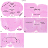
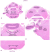
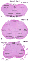
This review article is designed to serve as an introductory guide in neuroanatomy for toxicologic pathologists evaluating general toxicity studies. The article provides an overview of approximately 50 neuroanatomical subsites and their functional significance across 7 transverse sections of the brain. Also reviewed are 3 sections of the spinal cord, cranial and peripheral nerves (trigeminal and sciatic, respectively), and intestinal autonomic ganglia. The review is limited to the evaluation of hematoxylin and eosin-stained tissue sections, as light microscopic evaluation of these sections is an integral part of the first-tier toxicity screening of environmental chemicals, drugs, and other agents. Prominent neuroanatomical sites associated with major neurological disorders are noted. This guide, when used in conjunction with detailed neuroanatomic atlases, may aid in an understanding of the significance of functional neuroanatomy, thereby improving the characterization of neurotoxicity in general toxicity and safety evaluation studies.
Keywords: NTP; brain; functional neuroanatomy.; nerve; neuroanatomy; neuropathology; spinal cord.
Figures


Similar articles
-
STP position paper: Recommended practices for sampling and processing the nervous system (brain, spinal cord, nerve, and eye) during nonclinical general toxicity studies.Toxicol Pathol. 2013;41(7):1028-48. doi: 10.1177/0192623312474865. Epub 2013 Mar 7. Toxicol Pathol. 2013. PMID: 23475559
-
Histopathological evaluation of the nervous system in National Toxicology Program rodent studies: a modified approach.Toxicol Pathol. 2011 Apr;39(3):463-70. doi: 10.1177/0192623311401044. Epub 2011 Mar 23. Toxicol Pathol. 2011. PMID: 21430177
-
Society of toxicologic pathology position paper: review series: assessment of circulating hormones in nonclinical toxicity studies: general concepts and considerations.Toxicol Pathol. 2012 Aug;40(6):943-50. doi: 10.1177/0192623312444622. Epub 2012 May 8. Toxicol Pathol. 2012. PMID: 22569585 Review.
-
Morphologic aspects of rodent cardiotoxicity in a retrospective evaluation of National Toxicology Program studies.Toxicol Pathol. 2011 Aug;39(5):850-60. doi: 10.1177/0192623311413788. Epub 2011 Jul 11. Toxicol Pathol. 2011. PMID: 21747121
-
Society of Toxicologic Pathology position paper on best practices on recovery studies: the role of the anatomic pathologist.Toxicol Pathol. 2013;41(8):1159-69. doi: 10.1177/0192623313481513. Epub 2013 Mar 26. Toxicol Pathol. 2013. PMID: 23531793 Review.
Cited by
-
MALDI Imaging Mass Spectrometry Visualizes the Distribution of Antidepressant Duloxetine and Its Major Metabolites in Mouse Brain, Liver, Kidney, and Spleen Tissues.Drug Metab Dispos. 2024 Jun 17;52(7):673-680. doi: 10.1124/dmd.124.001719. Drug Metab Dispos. 2024. PMID: 38658163
-
Bisphenol-A Neurotoxic Effects on Basal Forebrain Cholinergic Neurons In Vitro and In Vivo.Biology (Basel). 2023 May 28;12(6):782. doi: 10.3390/biology12060782. Biology (Basel). 2023. PMID: 37372067 Free PMC article.
-
Biodistribution Analysis of an Anti-EGFR Antibody in the Rat Brain: Validation of CSF Microcirculation as a Viable Pathway to Circumvent the Blood-Brain Barrier for Drug Delivery.Pharmaceutics. 2022 Jul 12;14(7):1441. doi: 10.3390/pharmaceutics14071441. Pharmaceutics. 2022. PMID: 35890344 Free PMC article.
-
Research-Relevant Conditions and Pathology of Laboratory Mice, Rats, Gerbils, Guinea Pigs, Hamsters, Naked Mole Rats, and Rabbits.ILAR J. 2021 Dec 31;62(1-2):77-132. doi: 10.1093/ilar/ilab022. ILAR J. 2021. PMID: 34979559 Free PMC article. Review.
-
Host genetic diversity drives variable central nervous system lesion distribution in chronic phase of Theiler's Murine Encephalomyelitis Virus (TMEV) infection.PLoS One. 2021 Aug 20;16(8):e0256370. doi: 10.1371/journal.pone.0256370. eCollection 2021. PLoS One. 2021. PMID: 34415947 Free PMC article.
References
-
- Abdo KM, Wenk ML, Harry GJ, Mahler J, Goehl TJ, Irwin RD. Glycine modulates the toxicity of benzyl acetate in F344 rats. Toxicol Pathol. 1998;26:395–402. - PubMed
-
- Alves GS, Karakaya T, Fusser F, Kordulla M, O’Dwyer L, Christl J, Magerkurth J, Oertel-Knochel V, Knochel C, Prvulovic D, Jurcoane A, Laks J, Engelhardt E, Hampel H, Pantel J. Association of microstructural white matter abnormalities with cognitive dysfunction in geriatric patients with major depression. Psychiatry Res. 2012;203:194–200. - PubMed
-
- Andres KH, von During M, Veh RW. Subnuclear organization of the rat habenular complexes. J Comp Neurol. 1999;407:130–50. - PubMed
-
- Andrew-Jones L, Bradley A, Butt M, Garman B, Gumprecht L, Jensen K, McKay J, Pardo I, Radovsky A, TRoulois A, Sills R, Switzer R. Results from a survey of current practices for sampling of nervous system in rodents and non-rodents in general toxicity studies. STP/IFSTP Joint Symposium; Chicago. 2010.
Publication types
MeSH terms
Grants and funding
LinkOut - more resources
Full Text Sources
Other Literature Sources


