iBet uBet web content aggregator. Adding the entire web to your favor.


Link to original content: https://pubmed.ncbi.nlm.nih.gov/23773234
Evolutionary implications of the divergent long bone histologies of Nothosaurus and Pistosaurus (Sauropterygia, Triassic) - PubMed Skip to main page content
U.S. flag

An official website of the United States government

Dot gov

The .gov means it’s official.
Federal government websites often end in .gov or .mil. Before sharing sensitive information, make sure you’re on a federal government site.

Https

The site is secure.
The https:// ensures that you are connecting to the official website and that any information you provide is encrypted and transmitted securely.

Access keys NCBI Homepage MyNCBI Homepage Main Content Main Navigation
. 2013 Jun 18:13:123.
doi: 10.1186/1471-2148-13-123.

Evolutionary implications of the divergent long bone histologies of Nothosaurus and Pistosaurus (Sauropterygia, Triassic)

Affiliations

Evolutionary implications of the divergent long bone histologies of Nothosaurus and Pistosaurus (Sauropterygia, Triassic)

Anna Krahl et al. BMC Evol Biol. .

Abstract

Background: Eosauropterygians consist of two major clades, the Nothosauroidea of the Tethysian Middle Triassic (e.g., Nothosaurus) and the Pistosauroidea. The Pistosauroidea include rare Triassic forms (Pistosauridae) and the Plesiosauria of the Jurassic and Cretaceous. Long bones of Nothosaurus and Pistosaurus from the Muschelkalk (Middle Triassic) of Germany and France and a femur of the Lower Jurassic Plesiosaurus dolichodeirus were studied histologically and microanatomically to understand the evolution of locomotory adaptations, patterns of growth and life history in these two lineages.

Results: We found that the cortex of adult Nothosaurus long bones consists of lamellar zonal bone. Large Upper Muschelkalk humeri of large-bodied Nothosaurus mirabilis and N. giganteus differ from the small Lower Muschelkalk (Nothosaurus marchicus/N. winterswijkensis) humeri by a striking microanatomical specialization for aquatic tetrapods: the medullary cavity is much enlarged and the cortex is reduced to a few millimeters in thickness. Unexpectedly, the humeri of Pistosaurus consist of continuously deposited, radially vascularized fibrolamellar bone tissue like in the Plesiosaurus sample. Plesiosaurus shows intense Haversian remodeling, which has never been described in Triassic sauropterygians.

Conclusions: The generally lamellar zonal bone tissue of nothosaur long bones indicates a low growth rate and suggests a low basal metabolic rate. The large triangular cross section of large-bodied Nothosaurus from the Upper Muschelkalk with their large medullary region evolved to withstand high bending loads. Nothosaurus humerus morphology and microanatomy indicates the evolution of paraxial front limb propulsion in the Middle Triassic, well before its convergent evolution in the Plesiosauria in the latest Triassic. Fibrolamellar bone tissue, as found in Pistosaurus and Plesiosaurus, suggests a high growth rate and basal metabolic rate. The presence of fibrolamellar bone tissue in Pistosaurus suggests that these features had already evolved in the Pistosauroidea by the Middle Triassic, well before the plesiosaurs radiated. Together with a relatively large body size, a high basal metabolic rate probably was the key to the invasion of the Pistosauroidea of the pelagic habitat in the Middle Triassic and the success of the Plesiosauria in the Jurassic and Cretaceous.


Figures

Figure 1
Figure 1
Contrasting phylogenetic hypotheses of stem-group Sauropterygia and relationships of Nothosaurus. A, In the analysis of Holmes et al. [6], the putative pachypleurosaur Keichosaurus plots as a basal nothosauroid, and Pachypleurosauria and Nothosauroidea form a monophyletic taxon, resulting in the loss of the node Eusauropterygia; B, The classical view [4] with the Pachypleurosauria as the sister-group of Eusauropterygia including Nothosauroidea and Pistosauroidea; C, Phylogeny of the Nothosauridae [modified from 16] with the sampled species indicated in bold. Nothosaurus winterswijkensis and N. marchicus are more basal species than N. giganteus. N. mirabilis is one of the most derived nothosaur species.
Figure 2
Figure 2
Sauropterygian long bones with planes of section marked (arrows). A, humerus of Nothosaurus sp. indet. [from [93]]. Note that Nothosaurus humeri are morphologically more differentiated than those of the more advanced taxa. B, femur of Nothosaurus sp. indet. [from [2]]; C, humerus of Pistosaurus longaevus [from [8]]; D, femur of Pistosaurus longaevus [from [2]]; E, humerus of a plesiosaur [from [94]], figured for better morphological comparison with other sauropterygian long bones; F, femur of a plesiosaur [from [94]]. Proximal is at the top in all bones. Bones in A and D-F are shown in dorsal view; bones in B and C are shown in ventral view. Not to scale.
Figure 3
Figure 3
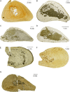
Overview photographs of Nothosaurus humeri thin sections in normal light. A-D and F-G, diaphyseal sections; E, metaphyseal thin section. In all images, ventral is at the bottom. A, N. marchicus/N. winterswijkensis humerus (MfN R 174–2); B, N. mirabilis humerus (SIPB R 54/2); C-E, N. giganteus humeri (C, SIPB R 53, D, SIPB R 45, E, MHI 1903, Section 1); F-G, Nothosaurus. sp. indet. humeri (F, MHI 1906, G, MHI 633). Note the triangular cross sections of the Nothosaurus humeri (A-D, F-G). Humeri in A and F are osteosclerotic (they have a low MI and the medullary regions are mostly infilled by endosteal bone and calcified cartilage), unlike the humeri in B, C and D which show thinner cortices and large medullary regions. The juvenile humerus in G has a small medullary cavity but highly vascularized cortical bone.
Figure 4
Figure 4
Overview photographs of Nothosaurus femur thin sections in normal light. A-D, diaphyseal sections. In all images, ventral is at the bottom. A, N. giganteus femur (SIPB R 49); B-C, Nothosaurus mirabilis femora (SIPB R 54/1, SIPB R 50/2); D, Nothosaurus sp. indet. femur (SMNS 84856). In contrast to the triangular Nothosaurus humeri, femora have approximately round cross sections. In the femora in A and C, strong remodeling and a tendency for expansion of the medullary region are visible. The femur in D is clearly osteosclerotic, and the one in B also shows some osteosclerosis.
Figure 5
Figure 5
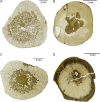
Overview photographs of long bone thin sections in normal light of Pistosaurus and Plesiosaurus. A-D, diaphyseal sections; A-B, Pistosaurus humeri (A, SIPB R 46, B, SMNS 84825); C, Pistosaurus femur (SIPB R 74); D, Plesiosaurus humerus SIPB R 90). In all images, ventral is at the bottom. The Pistosaurus humeri in A and B have a thick cortex and a very small medullary region unlike the Pistosaurus femur in C, which resembles the nothosaur femur in Figure 4C. The Plesiosaurus femur in D exhibits incipient osteoporosis, typical for pelagically adapted marine tetrapods.
Figure 6
Figure 6
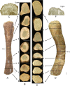
Polished serial cross sections and epiphyseal longitudinal sections of Nothosaurus mirabilis long bones in comparison. A, left femur (SIPB R 54/1) in dorsal view, anterior is to the right, proximal is to the top. D, left humerus (SIPB R 54/2) in dorsal view, anterior is to the right, proximal is to the top. Arrows in A and D assign the corresponding polished section to each section plane (marked by a dotted line). Serial transverse sections of femur (B 1–8) and of humerus (E 1–9) are numbered and described in the text from mid-diaphysis to proximal end and distal end, respectively. In all serial sections, anterior is to the right and dorsal side is up. Of B 1 and E 1 only mid-diaphyseal thin sections were produced which are pictured in Figure 3B and Figure 4B; C, longitudinal thin section of the proximal femur epiphysis. Plane of section is in anteroposterior direction. F, longitudinal thin section of the proximal humerus epiphysis. Plane of section is in dorsoventral direction. Femur sections B 1–8 display the generalized tetrapod long bone microanatomy. The cortex becomes thinner, and the amounts of secondary cancellous bone increase from the middle region of the shaft (B 1) towards both epiphyses (B 4 and B 8). The open medullar space enlarges from mid-diaphysis towards both articular ends and extends to the spongy endochondral and endosteal bone which provides the base for the articular cartilage (C). Humerus sections E 1–9 follow the same pattern as B 1–8, but throughout the entire humerus the cortex is much thinner, and secondary cancellous bone and endochodral and endosteal bone are only sparsely present. The medullary region extends proximally and distally to a very thin bony layer onto which the articular cartilage was placed in the living animal (F). Scale bars equal 10 mm.
Figure 7
Figure 7
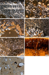
Long bone histology of species of Nothosaurus. A-D, thin sections in diaphysial region; E, F, thin sections in metaphyseal region; A-C, E, in polarized light; D, F, in normal light. A, humerus of N. marchicus/N. winterswijkensi (MfN R 174–2) showing a cortex which consists of well vascularized LZB. There is a thin, highly birefringent circumferential layer of lamellar bone that separates the medullary region from cortical bone. Trabeculae of endosteal bone with a core of calcified cartilage partially fill in the medullary region; B, humerus of N. giganteus (SIPB R 40), showing Sharpey’s fibres in cortical LZB of the posterior sector of the cross section. Note how the fibers influence the appositional bone organization, creating a radial appearance of the cortical bone, and how the vascular canals are deflected radially. Detail of humerus of N. giganteus (SIPB R 53) in polarized (C) and normal light (D). C, the outermost cortex of this specimen is composed almost entirely of thin layers of lamellar bone; D, closely spaced LAGs (EFS) in the outermost cortex of a humeral cross section indicate that diaphyseal growth has nearly completely ceased. E, humerus of N. giganteus (MHI 1903), Section 2 (the relatively more distal one), has a cortex composed of RLLZB showing regularly spaced zones, annuli and LAGs. Arrows mark the visible LAGs. F, humerus of N. giganteus (MHI 1903), Section 1 (the relatively more proximal one), the highly vascularized lamellar zonal cortex merges towards the shaft into cyclial LZB as seen in E. Abbreviations: cc, calcified cartilage, eb, endosteal bone, emb, embryonic bone, sf, Sharpey’s fibers.
Figure 8
Figure 8
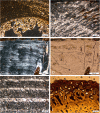
Long bone histology of Nothosaurus species. A-G, thin sections in diaphysial region; A-E, in polarized light; F, G, in normal light. A, humerus of N. mirabilis (SIPB R 54/2), cortex with radial vascular canals with funnel structures. Radial vascular canals undulate and seem to follow the orientation of the Sharpey’s fibers; B, medullary region of femur of N. mirabilis (SIPB R 54/2), showing secondary cancellous bone in the medullary region, consisting of several generations of endosteal lamellar bone; C, inner cortex and medullary region of femur of N. mirabilis (SIPB R 50/1), showing well vascularized LZB. Thick endosteal trabeculae line the medullary region. Large erosion lacunae are visible in deeper cortex; D, inner cortex and medullary region of humerus of Nothosaurus sp. indet. (MHI 1906). Embryonic bone is preserved up to the hatching or birth line (marked by the arrow). The remainder of the cortex is made up of highly vascularized LZB. Towards the center of the bone, a highly birefringent circumferential layer separates the cortical bone from the medullary region. It has partially been resorbed. In the medullary region, there are trabeculae consisting of endosteal bone and a core of calcified cartilage. E, humerus of Nothosaurus sp. indet. (MHI 633). Deposition of the cortex began with embryonic bone up to hatching or birth line (marked by the arrow). Cortical bone consists otherwise of highly vascularized incipient FLB; F, humerus of an adult individual of Nothosaurus sp. indet. (SMNS 84856). LAGs in the cortex (marked by arrows) become more closely spaced towards the bone surface, but there is no EFS in the outermost cortex; G, Endosteal lamellar bone surrounding cores of calcified cartilage in the medullary region of MfN R 174–2. Abbreviations: cc, calcified cartilage, eb, endosteal bone, emb, embryonic bone, fs, funnel structures, sf, Sharpey’s fibres.
Figure 9
Figure 9
Micrographs of longitudinal thin sections of Nothosaurus mirabilis long bones. A, proximal epiphysis of N. mirabilis humerus (SIPB R 54/2) in polarized light. The section shows the growth plate below the articular cartilage. The endochondral bone is very thin at the proximal epiphysis, and there are only a few endochondral trabeculae, which have mostly been coated by endosteal bone. B-E, proximal epiphysis of N. mirabilis femur (SIPB R 54/1). B and C show the growth plate below the articular cartilage in normal transmitted and in polarized light. The trabeculae of the growth plate consist of cartilage covered by endochondral bone. D-E, trabeculae in deeper cortical regions of the femur in normal transmitted light (D) and polarized light (E). The trabeculae still have a calcified cartilage core but were subject to remodeling activity. Abbreviations: eb, endosteal bone.
Figure 10
Figure 10
Comparison of the humerus histology of two Pistosaurus longaevus individuals. Match the juvenile SIPB R 46 (A, C) and the adult SMNS 84825 (B, D). A-B, outer cortex in normal transmitted light. Radially and longitudinally vascularized FLB is interrupted by several thick parallel-fibred annuli (marked by arrows). Vascular spaces are filled in by more highly organized bone tissue to a greater degree in the older individual (B) than in the juvenile (A). C, D, inner cortex in polarized light. Embryonic bone is preserved in both individuals and extends to the hatching or birth line (marked by arrow). The rest of the cortex consists of strongly vascularized FLB with distinctly radial primary osteons. Although the medullary region is lined by circumferential layers of endosteal bone in both specimens, the cortex of the juvenile (C) is more highly vascularized than that of the older one (D). E-G, detail (box in C) of the FLB of the juvenile (SIPB R 46) showing the framework of woven-fibered bone, which is only incompletely filled in by lamellar bone. The primary osteons are thus still incompletely formed. The same view is seen in normal-transmitted light in E, in polarized light in F, and with the gypsum plate in G. Abbreviations: emb, embryonic bone, fb, fibrous or woven-fibered bone, lb, lamellar bone, po, primary osteon.
Figure 11
Figure 11
Bone histology of Pistosaurus longaevus and Plesiosaurus dolichodeirus. A and B, Pistosaurus longaevus femur (SIPB R 74), seen in normal-transmitted (A) and polarized light (B). C and D, Plesiosaurus dolichodeirus femur (SIPB R 90) seen in normal-transmitted light (C) and with gypsum plate (D). A and B, detail of the lower right sector in cross section in Figure 5C. Unlike the humeri, the cortex of the femur consists of LZB except for the deepest cortical regions. The detail shows a growth cycle that locally consists of radial FLB following one of LZB. C, cortical bone composed of FLB of Plesiosaurus dolichodeirus (SIPB R 90). The primary cortex was subject to Haversian remodeling, partially obscuring it. An EFS is observable. Note the relatively small primary vascular canals and the large secondary osteons. D, detail of SIPB R 90, showing the radial fabric of primary bone, which appears to have been inducing resorption activity that led to the formation of secondary osteons. Abbreviations: pc, primary vascular canal, rflb, radial fibrolamellar bone, so, secondary osteon.
Figure 12
Figure 12
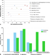
Variation of medullary index. A, Scatter plot of medullary index (MI) vs. bone circumference of all sampled sauropterygian long bones, subdivided by genus, species and long bone type. Three clusters can be distinguished, reflecting either very small, intermediate, or large medullary regions. Note the trend of increasing MI with increasing body size in the genus Nothosaurus. Pistosaurus differs from the other sauropterygians in its very small medullary region in the humerus. B, comparison of the MI for each bone type of each species. The highest MI is seen in the long bones of Nothosaurus giganteus. Also note the great differences between the MI of the humeri and the femur of Pistosaurus.

Similar articles

Cited by

References

    1. Benson RBJ, Evans M, Druckenmiller PS. High diversity, low disparity and small body size in plesiosaurs (Reptilia, Sauropterygia) from the Triassic-Jurassic boundary. PLoS One. 2012;7:1–15. - PMC - PubMed
    1. Meyer H, der Vorwelt Zur Fauna. Die Saurier des Muschelkalkes mit Rücksicht auf die Saurier aus buntem Sandstein und Keuper. Frankfurt am Main: Heinrich Keller; 1847-1855.
    1. Rieppel O. Osteology of Simosaurus gaillardoti, and the phylogenetic interrelationships of stem-group Sauropterygia. Field Geol, N S. 1994;28:1–85.
    1. Rieppel O. In: In Encyclopedia of Paleoherpethology. Kuhn PWO, editor. München: Verlag Dr. Friedrich Pfeil; 2000. Sauropterygia I; pp. 1–134.
    1. Liu J, Rieppel O, Jiang D-Y, Aitchison JC, Motani R, Zhang Q-Y, Zhou C-Y, Sun Y-Y. A new pachypleurosaur (Reptilia: Sauropterygia) from the lower Middle Triassic of southwestern China and the phylogenetic relationships of Chinese pachypleurosaurs. J Vert Paleont. 2011;31:292–302. doi: 10.1080/02724634.2011.550363. - DOI

LinkOut - more resources