Expression dynamics and protein localization of rhabdomeric opsins in Platynereis larvae
- PMID: 23667045
- PMCID: PMC3687135
- DOI: 10.1093/icb/ict046
Expression dynamics and protein localization of rhabdomeric opsins in Platynereis larvae
Abstract
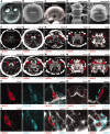
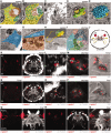
The larval stages of polychaete annelids are often responsive to light and can possess one to six eyes. The early trochophore larvae of the errant annelid Platynereis dumerilii have a single pair of ventral eyespots, whereas older nectochaete larvae have an additional two pairs of dorsal eyes that will develop into the adult eyes. Early Platynereis trochophores show robust positive phototaxis starting on the first day of development. Even though the mechanism of phototaxis in Platynereis early trochophore larvae is well understood, no photopigment (opsin) expression has yet been described in this stage. In late trochophore larvae, a rhabdomeric-type opsin, r-opsin1, expressed in both the eyespots and the adult eyes has already been reported. Here, we identify another Platynereis rhabdomeric opsin, r-opsin3, that is expressed in a single photoreceptor in the eyespots in early trochophores, suggesting that it mediates early larval phototaxis. We also show that r-opsin1 and r-opsin3 are expressed in adjacent photoreceptor cells in the eyespots in later stages, indicating that a second eyespot-photoreceptor differentiates in late trochophore larvae. Using serial transmission electron microscopy (TEM), we identified and reconstructed both photoreceptors and a pigment cell in the late larval eyespot. We also characterized opsin expression in the adult eyes and found that the two opsins co-express there in several photoreceptor cells. Using antibodies recognizing r-opsin1 and r-opsin3 proteins, we demonstrate that both opsins localize to the rhabdomere in all six eyes. In addition, we found that r-opsin1 mRNA is localized to, and translated in, the projections of the adult eyes. The specific changes we describe in opsin transcription and translation and in the cellular complement suggest that the six larval eyes undergo spectral and functional maturation during the early planktonic phase of the Platynereis life cycle.
Figures


Similar articles
-
Spectral Tuning of Phototaxis by a Go-Opsin in the Rhabdomeric Eyes of Platynereis.Curr Biol. 2015 Aug 31;25(17):2265-71. doi: 10.1016/j.cub.2015.07.017. Epub 2015 Aug 6. Curr Biol. 2015. PMID: 26255845
-
CRISPR/CAS9 mutagenesis of a single r-opsin gene blocks phototaxis in a marine larva.Proc Biol Sci. 2019 Jun 12;286(1904):20182491. doi: 10.1098/rspb.2018.2491. Epub 2019 Jun 5. Proc Biol Sci. 2019. PMID: 31161907 Free PMC article.
-
Stable transgenesis in the marine annelid Platynereis dumerilii sheds new light on photoreceptor evolution.Proc Natl Acad Sci U S A. 2013 Jan 2;110(1):193-8. doi: 10.1073/pnas.1209657109. Epub 2013 Jan 2. Proc Natl Acad Sci U S A. 2013. PMID: 23284166 Free PMC article.
-
Phototaxis and the origin of visual eyes.Philos Trans R Soc Lond B Biol Sci. 2016 Jan 5;371(1685):20150042. doi: 10.1098/rstb.2015.0042. Philos Trans R Soc Lond B Biol Sci. 2016. PMID: 26598725 Free PMC article. Review.
-
Simple Eyes, Extraocular Photoreceptors and Opsins in the American Horseshoe Crab.Integr Comp Biol. 2016 Nov;56(5):809-819. doi: 10.1093/icb/icw093. Epub 2016 Jul 21. Integr Comp Biol. 2016. PMID: 27444526 Review.
Cited by
-
Elongation capacity of polyunsaturated fatty acids in the annelid Platynereis dumerilii.Open Biol. 2024 Jun;14(6):240069. doi: 10.1098/rsob.240069. Epub 2024 Jun 12. Open Biol. 2024. PMID: 38864244 Free PMC article.
-
Opsin expression varies across larval development and taxa in pteriomorphian bivalves.Front Neurosci. 2024 Mar 18;18:1357873. doi: 10.3389/fnins.2024.1357873. eCollection 2024. Front Neurosci. 2024. PMID: 38562306 Free PMC article.
-
Characterization of eyes, photoreceptors, and opsins in developmental stages of the arrow worm Spadella cephaloptera (Chaetognatha).J Exp Zool B Mol Dev Evol. 2023 Jul;340(5):342-353. doi: 10.1002/jez.b.23193. Epub 2023 Feb 28. J Exp Zool B Mol Dev Evol. 2023. PMID: 36855226 Free PMC article.
-
The diversity of invertebrate visual opsins spanning Protostomia, Deuterostomia, and Cnidaria.Dev Biol. 2022 Dec;492:187-199. doi: 10.1016/j.ydbio.2022.10.011. Epub 2022 Oct 19. Dev Biol. 2022. PMID: 36272560 Free PMC article. Review.
-
Molecular and cellular architecture of the larval sensory organ in the cnidarian Nematostella vectensis.Development. 2022 Aug 15;149(16):dev200833. doi: 10.1242/dev.200833. Epub 2022 Aug 24. Development. 2022. PMID: 36000354 Free PMC article.
References
-
- Arendt D. Evolution of eyes and photoreceptor cell types. Int J Dev Biol. 2003;47:563–71. - PubMed
-
- Arendt D. Ciliary photoreceptors with a vertebrate-type opsin in an invertebrate brain. Science. 2004;306:869–71. - PubMed
-
- Arendt D, Tessmar K, de Campos-Baptista M-IM, Dorresteijn A, Wittbrodt J. Development of pigment-cup eyes in the polychaete Platynereis dumerilii and evolutionary conservation of larval eyes in Bilateria. Development. 2002;129:1143–54. - PubMed
Publication types
MeSH terms
Substances
LinkOut - more resources
Full Text Sources
Other Literature Sources


