iBet uBet web content aggregator. Adding the entire web to your favor.


Link to original content: https://pubmed.ncbi.nlm.nih.gov/23502536
Local potentiation of excitatory synapses by serotonin and its alteration in rodent models of depression - PubMed Skip to main page content
U.S. flag

An official website of the United States government

Dot gov

The .gov means it’s official.
Federal government websites often end in .gov or .mil. Before sharing sensitive information, make sure you’re on a federal government site.

Https

The site is secure.
The https:// ensures that you are connecting to the official website and that any information you provide is encrypted and transmitted securely.

Access keys NCBI Homepage MyNCBI Homepage Main Content Main Navigation
. 2013 Apr;16(4):464-72.
doi: 10.1038/nn.3355. Epub 2013 Mar 17.

Local potentiation of excitatory synapses by serotonin and its alteration in rodent models of depression

Affiliations

Local potentiation of excitatory synapses by serotonin and its alteration in rodent models of depression

Xiang Cai et al. Nat Neurosci. 2013 Apr.

Abstract

The causes of major depression remain unknown. Antidepressants elevate concentrations of monoamines, particularly serotonin, but it remains uncertain which downstream events are critical to their therapeutic effects. We found that endogenous serotonin selectively potentiated excitatory synapses formed by the temporoammonic pathway with CA1 pyramidal cells via activation of serotonin receptors (5-HT(1B)Rs), without affecting nearby Schaffer collateral synapses. This potentiation was expressed postsynaptically by AMPA-type glutamate receptors and required calmodulin-dependent protein kinase-mediated phosphorylation of GluA1 subunits. Because they share common expression mechanisms, long-term potentiation and serotonin-induced potentiation occluded each other. Long-term consolidation of spatial learning, a function of temporoammonic-CA1 synapses, was enhanced by 5-HT(1B)R antagonists. Serotonin-induced potentiation was quantitatively and qualitatively altered in a rat model of depression, restored by chronic antidepressants, and required for the ability of chronic antidepressants to reverse stress-induced anhedonia. Changes in serotonin-mediated potentiation, and its recovery by antidepressants, implicate excitatory synapses as a locus of plasticity in depression.


Conflict of interest statement

The authors declare they have no conflicting financial interests.

Figures

Figure 1
Figure 1. 5-HT1BR activation selectively potentiates TA-CA1 cell excitatory synapses
(a) Promoting accumulation of endogenous serotonin by bath application of the tricyclic antidepressant imipramine (2 μM) potentiated TA-CA1 fEPSPs in SLM of area CA1 of acutely prepared hippocampal slices (n=8 slices) in control saline (black), but not in the slices pretreated with 5-HT1BR antagonist isamoltane (10 μM) (red; n=9 slices). Sample traces before (black) and 60 min after imipramine application (red) in control saline (upper row) or in the presence of isamoltane (lower row) are shown at right. (b) Group data showing the effect of antidepressants on TA-CA1 fEPSPs in control slices (fluoxetine, n=4 slices; citalopram, n=3 slices) and the effect of anpirtoline (50 μM) on TA-CA1 fEPSPs in control slices (red, n=11 slices; ANOVA F(3,20)=9.751, p<0.001) or slices pretreated for 60 min with antidepressants (n=5 slices pretreated with imipramine; n=5 slices with fluoxetine; n=3 slices with citalopram; F(3,20)=7.32, p=0.002). Bonferroni post-hoc tests revealed that isamoltane prevented the increase in fEPSP slope observed with antidepressants alone (p<0.05 vs. Imipramine, Fluoxetine, or Citalopram), and that anpirtoline treatment differed from each of these pretreatment+anpirtoline conditions (p<0.05). (c) The selective 5HT1BR agonist, anpirtoline, reversibly increased the slope of TA-CA1 fEPSPs in SLM of area CA1 (black circles; n=11 slices). Isamoltane (10 μM) blocked the effect of anpirtoline on TA-CA1 fEPSPs (red triangles; n=5 slices). (d) Anpirtoline selectively enhanced TA-CA1 EPSCs recorded in whole-cell voltage-clamp mode, but not simultaneously evoked SC-CA1 EPSCs in a two-pathway experimental design (n=9 cells). Sample traces are shown at right. The selective increase in TA-CA1 fEPSPs by serotonin, but not SC-CA1 fEPSPs, is consistent with the selective localization of 5-HT1BRs in SLM. *p<0.05, **p<0.01 compared with before anpirtoline or antidepressant; paired t-test.
Figure 2
Figure 2. Expression of 5-HT1BR-induced potentiation of TA-CA1 synapses is mediated postsynaptically
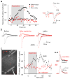
(a) Time course of changes of TA-CA1 fEPSP slope (black circles), fiber volley (black triangles), and paired-pulse ratio (PPR, red circles) before and after anpirtoline application in a typical experiment. Representative traces shown at right, with the fiber volley at higher resolution above. Neither fiber volley (t(6)=0.039, p=0.97) nor PPR (t(10)=1.17, p=0.27) changed with anpirtoline treatment. (b) Whole-cell voltage-clamp recordings of EPSCs at −70 and +40 mV in the same cell before (black) and 40 min after application of anpirtoline (red). (c) Left: Image of a CA1 cell filled with Alexa 594 from a somatic whole-cell pipette indicating sites of microphotolysis of caged glutamate on distal apical dendrites in SLM or oblique dendrites in SR of CA1 pyramidal cells. Scale bar = 20 μm. Right: bath application of anpirtoline increased photolysis-evoked EPSPs elicited from dendrites in SLM in acutely prepared hippocampal brain slices (n=5 cells) but not from dendrites in SR (n=6 cells in separate experiments), indicating that 5-HT1BR activation selectively enhanced postsynaptic glutamate responses at sites of TA synapse formation. Sample EPSP-like photolysis responses elicited from dendrites in SLM and SR before (black) and after anpirtoline application (red) are shown at right.
Figure 3
Figure 3. Anpirtoline activates CaMK and increases phosphorylation of GluA1 at serine 831 in SLM tissue
(a,b) Representative Western blots and quantification showing that anpirtoline (50 μM, 2 hr) time dependently increased phosphorylation of alpha and beta CaMK at T286 (molecular weights 52 and 60 kD; F(5,18)=4.651, p=0.007) and GluA1 at S831 (F(5,24)=3.706, p=0.013), but did not affect total GluA1 expression (F(5,18)=0.863, p=0.524) in isolated CA1 SLM tissue sections, subdissected from whole hippocampal slices. (c) The effect of anpirtoline on TA-CA1 fEPSPs was blocked by bath application of the specific CaMK inhibitor, KN62 (5 μM)(n=6 slices), but not by bath application of the cell-permeable PKA inhibitor, PKI 14-22 (1 μM)(n=3 slices). Intracellular dialysis of the CaMK inhibitor, autocamtide-2-related inhibitory peptide (AIP, 2 μM) via patch-pipettes (n=5 cells) blocked potentiation of TA-CA1 EPSPs recorded in whole-cell mode. (d) Anpirtoline increased phosphorylation of GluA1 at S831 in isolated CA1 SLM tissue sections but not at S845. (e) Anpirtoline (50 μM for 60 min) increased phosphorylation of CaMK T286 (left, Mann-Whitney U z=2.09, p=0.04) and GluA1 S831 (right, Mann-Whitney U z=2.09, p=0.04) in tissue punches taken from the core of the Nucleus Accumbens (n=6 slices each). (f) Anpirtoline did not potentiate TA-CA1 fEPSPs in slices from GluA1 S831A knock-in mice (n=7 slices from 4 animals), but produced strong potentiation in slices from wild-type littermate mice (n=4 slices from 4 animals). *, p<0.05; **, p<0.01 compared with before anpirtoline. Full-length blots are presented in Supplementary Figure 7.
Figure 4
Figure 4. 5-HT1BR-mediated potentiation occludes LTP at TA-CA1 synapses and influences memory consolidation
(a) After induction of LTP with high frequency stimulation (4 trains, 1 sec per train at 100 Hz, 5 min interval), bath application of anpirtoline (50 μM) failed to induce further potentiation of TA-CA1 fEPSP slope (n=8 slices). (b) The effect of anpirtoline on TA-CA1 fEPSP slope remained occluded after induction of LTP, even when the stimulation intensity was decreased to return the fEPSP to the baseline level (n=5 slices). Representative traces are shown below. Scale bar = 0.1 mV, 20 ms. (c) Anpirtoline-induced potentiation of TA-CA1 fEPSP slope occluded tetanus-induced LTP at TA-CA1 synapses (n=4 slices). (d) Anpirtoline neither enhanced SC-CA1 fEPSPs nor occluded LTP of SC-CA1 synapses (red; n=7 slices). LTP of SC-CA1 fEPSPs in control slices shown in black (n=5 slices). Only every other data point is plotted to allow the two data sets to be distinguished.
Figure 5
Figure 5. Spatial memory consolidation is affected by endogenous activation of 5-HT1BRs
Latency to find the target platform in the Morris water maze (a) decreased significantly between the first (naïve) and last training trial (trained), demonstrating that the rats learned the location of the platform; remained short during a probe trial administered 24 hrs after the last training trial, demonstrating that the rats remembered the platform location; and remained significantly shorter than naïve when tested 28 days after the final training trial, demonstrating memory consolidation. Rats administered a 5-HT1BR antagonist (SB216641, 4 mg/kg, i.p.) daily for 14 days starting after the 24 hr probe trial displayed a significantly shorter escape latency in the consolidation trial than the control group (0.9% NaCl, i.p.). Representative examples of the swim path in the maze from the start (white) to stop (black) positions are shown at left for control and SB-treated individuals. SB-treated animals also spent more time in the target quadrant during the 28 day consolidation probe trial than controls (b), and swam a significantly shorter distance than controls (c). Both groups had identical swim speeds (c), however, indicating that the difference in performance was not due to altered motor function. Full swim paths for two different individuals in the probe trials are shown at left. Dashed line indicates random performance. **, p<0.05; *, p=0.06; n= 8 animals per group; post-hoc t-test. Further statistical information can be found in the Methods.
Figure 6
Figure 6. Chronic stress enhances, whereas chronic antidepressants eliminate, the effect of anpirtoline on TA-CA1 synaptic transmission
(a) Changes in TA-CA1 EPSP slope in slices from CUS animals (red, n=11 cells) and control littermates (black, n=10 cells) showing the significantly larger amplitude and persistence of potentiation in CUS animals, compared to controls. Representative traces before and after anpirtoline application and after 60 min of washout shown at right. Scale bars = 2 mV, 50 ms. (b) Changes in TA-CA1 EPSP slope induced by anpirtoline in slices from CUS animals (red) are larger and more persistent than the potentiation seen in slices from controls (black)(controls: peak change in fEPSP slope = 162±3%, fEPSP slope after 60 min wash = 106±3%; CUS: peak change = 226±23%, 218±21% after 60 min wash)(n=10,11 slices). (c) Anpirtoline produced no significant effect on fEPSP slope in slices from animals treated for 21–28 days with antidepressant imipramine (100 mg/L, red) or fluoxetine (80 mg/L, blue), whereas it doubled fEPSP slope in slices from controls in interleaved experiments (black) (imipramine: n=9 slices; fluoxetine: n=5 slices; controls: n=7 slices). (d) Administration of fluoxetine for the final 3 weeks in animals subjected to six weeks of CUS (blue, n=12 slices) resulted in a restoration of transient anpirtoline-induced potentiation (black, n=11 slices), clearly different from the irreversible potentiation in CUS animals (red, n=5 slices).
Figure 7
Figure 7. 5-HT1BR-dependent potentiation of GluA1 receptors is necessary for the therapeutic action of antidepressants
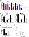
(a) Sucrose preference was high in vehicle-treated control C57BL6j mice (black) and remained high. SDS eliminated sucrose preference after one week (light blue). Fluoxetine (80 mg/l) restored sucrose preference in SDS animals in which it had been lost (red). The 5-HT1BR antagonist SB224289 (4 mg/liter in drinking water) had no effect on sucrose preference in non-SDS mice (gray) but prevented restoration of sucrose preference in SDS mice administered fluoxetine (dark blue). * = significantly less than corresponding baseline. (b) Sucrose preference was high in naïve 5-HT1BR−/−mice (blue) and wild-type mice (Sv129Imj, black), and was decreased in both strains after CUS. Fluoxetine restored sucrose preference in wild-type, but not 5-HT1BR−/− mice. * = significantly different than baseline in same genotype (Bonferroni corrected post-hoc p<0.05). § = significantly different than after CUS (Bonferroni corrected post-hoc p<0.05). We conclude that 5-HT1BR activation is thus required for fluoxetine to exert its therapeutic action. (c) CUS decreased sucrose preference in control C57BL6j mice (Bonferroni corrected post-hoc p<0.02), which was restored by fluoxetine [baseline vs. fluoxetine p>0.5]. Sucrose preference was lower in GluA1 S831A mice (red) than in wild-type mice (C57BL6j, black)[Student’s t(19)= 3.37, p<0.005]. Sucrose preference in GluA1 S831A mice was not affected by fluoxetine [t(14)= 0.335, p>0.7 with paired t-test], and antidepressant treated GluA1 S831A mice had lower sucrose preference than controls [Student’s t(25)=2.42, p<0.05 with Bonferroni adjustment for multiple comparisons]. * = significantly less than C57BL6j baseline, †, significantly different than C57BL6j baseline, but not GluA1 S831A baseline. (d) In the tail suspension test, C57BL6J littermates (black) treated with imipramine spent less time immobile than wild-types treated with saline [t(10)= 4.10, p<0.005]. S813A mice (red) treated with imipramine spent less time immobile than GluA1 S831A mice treated with saline [t(13)= 2.35, p<0.05]. * = significantly different than control in same genotype. A behavioral response to acute administration of antidepressants was thus preserved in this assay, unlike the response to chronic antidepressants in the sucrose preference test. (e) Latency to feeding in the novelty-suppressed feeding test was longer in GluA1 S831A mice (n=14) than in wild-type C57BL6j mice (n=13)[t(27)= −2.10, p<0.05]. GluA1 S831A mice did not differ from wild-type littermates with regard to time spent in the center in the open field test, but completed fewer line crossings than littermates, consistent with a hypo-locomotor phenotype, but not increased anxiety [t(13)= −2.29, p<0.05]. * = significantly different than wildtype. Taken together with lower sucrose preference, GluA1 S831A mice display a depressive-like behavioral phenotype. Further statistical information can be found in the Methods.

Similar articles

Cited by

References

    1. Cai X, Liang CW, Muralidharan S, Kao JPY, Tang CM, Thompson SM. Unique roles of SK and Kv4.2 potassium channels in dendritic integration. Neuron. 2004;44:351–364. - PubMed
    1. Dewar KM, Grondin L, Carli M, Lima L, Reader TA. [3H]paroxetine binding and serotonin content of rat cortical areas, hippocampus, neostriatum, ventral mesencephalic tegmentum, and midbrain raphe nuclei region following p-chlorophenylalanine and p-chloroamphetamine treatment. J Neurochem. 1992;58:250–257. - PubMed
    1. Remondes M, Schuman EM. Role for a cortical input to hippocampal area CA1 in the consolidation of a long-term memory. Nature. 2004;431:699–703. - PubMed
    1. Santarelli L, Saxe M, Gross C, Surget A, Battaglia F, Dulawa S, Weisstaub N, Lee J, Duman R, Arancio O, Belzung C, Hen R. Requirement of hippocampal neurogenesis for the behavioral effects of antidepressants. Science. 2003;301:805–809. - PubMed

Reference List

    1. Krishnan V, Nestler EJ. The molecular neurobiology of depression. Nature. 2008;455:894–902. - PMC - PubMed
    1. Billings AG, Cronkite RC, Moos RH. Social-environmental factors in unipolar depression: comparisons of depressed patients and nondepressed controls. J Abnorm Psychol. 1983;92:119–133. - PubMed
    1. Heninger GR, Delgado PL, Charney DS. The revised monoamine theory of depression: a modulatory role for monoamines, based on new findings from monoamine depletion experiments in humans. Pharmacopsychiatry. 1996;29:2–11. - PubMed
    1. Duman RS, Aghajanian GK. Synaptic dysfunction in depression: potential therapeutic targets. Science. 2012;338:68–72. - PMC - PubMed
    1. Sanacora G, Zarate CA, Krystal JH, Manji HK. Targeting the glutamatergic system to develop novel, improved therapeutics for mood disorders. Nat Rev Drug Discov. 2008;7:426–437. - PMC - PubMed

Publication types

MeSH terms