iBet uBet web content aggregator. Adding the entire web to your favor.


Link to original content: https://pubmed.ncbi.nlm.nih.gov/23452092
Selective degeneration of dopaminergic neurons by MPP(+) and its rescue by D2 autoreceptors in Drosophila primary culture - PubMed Skip to main page content
U.S. flag

An official website of the United States government

Dot gov

The .gov means it’s official.
Federal government websites often end in .gov or .mil. Before sharing sensitive information, make sure you’re on a federal government site.

Https

The site is secure.
The https:// ensures that you are connecting to the official website and that any information you provide is encrypted and transmitted securely.

Access keys NCBI Homepage MyNCBI Homepage Main Content Main Navigation
. 2013 Aug;126(4):529-40.
doi: 10.1111/jnc.12228. Epub 2013 Mar 24.

Selective degeneration of dopaminergic neurons by MPP(+) and its rescue by D2 autoreceptors in Drosophila primary culture

Affiliations

Selective degeneration of dopaminergic neurons by MPP(+) and its rescue by D2 autoreceptors in Drosophila primary culture

Lyle Wiemerslage et al. J Neurochem. 2013 Aug.

Abstract

Drosophila melanogaster is widely used to study genetic factors causing Parkinson's disease (PD) largely because of the use of sophisticated genetic approaches and the presence of a high conservation of gene sequence/function between Drosophila and mammals. However, in Drosophila, little has been done to study the environmental factors which cause over 90% of PD cases. We used Drosophila primary neuronal culture to study degenerative effects of a well-known PD toxin MPP(+) . Dopaminergic (DA) neurons were selectively degenerated by MPP(+) , whereas cholinergic and GABAergic neurons were not affected. This DA neuronal loss was because of post-mitotic degeneration, not by inhibition of DA neuronal differentiation. We also found that MPP(+) -mediated neurodegeneration was rescued by D2 agonists quinpirole and bromocriptine. This rescue was through activation of Drosophila D2 receptor DD2R, as D2 agonists failed to rescue MPP(+) -toxicity in neuronal cultures prepared from both a DD2R deficiency line and a transgenic line pan-neuronally expressing DD2R RNAi. Furthermore, DD2R autoreceptors in DA neurons played a critical role in the rescue. When DD2R RNAi was expressed only in DA neurons, MPP(+) toxicity was not rescued by D2 agonists. Our study also showed that rescue of DA neurodegeneration by Drosophila DD2R activation was mediated through suppression of action potentials in DA neurons.

Keywords: D2 agonist; DD2R; Drosophila melanogaster; Parkinson's disease; primary neuronal culture.


Conflict of interest statement

Conflict of Interest: Authors declare that they have no conflicts of interest.

Figures

Figure 1
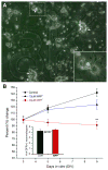
Figure 1. MPP+ reduces number of dopaminergic neurons in Drosophila primary culture
(A) An overlapped image of bright-field and GFP signals in a marked square (8K in this case). Six GFP(+) dopaminergic (DA) neurons are indicated by arrows. Neuronal cultures were derived from a cross between TH-Gal4 and UAS-GFP lines (=TH-GFP). As shown previously (Park et al, 2007), these cultures were plated onto photoetched coverslips which allow for identifying a square containing GFP(+) neurons that can then be returned to at a later time. Scale bar = 20μm. Inset An enlarged view of the dotted square in (A). showing a GFP(+) neuron is in a neuronal cluster containing GFP(−) neurons. (B) A graph showing percent change in number of GFP(+) DA neurons in the absence and presence of MPP+. Number of GFP(+) neurons at 5 and 9 days in vitro (DIV) was normalized to that at 3 DIV. The control and MPP+ treated cultures have a similar number of GFP(+) DA neurons at 3DIV (Inset, see below). However, at 9 DIV, the number of GFP(+) neurons in the control dish has increased while the number has slightly decreased in the MPP+ treated cultures. Inset A graph showing number of GFP(+) neurons per each square averaged at 3 days in vitro (DIV). Coverslips were randomly split into three groups: control, 10μM MPP+ or 40μM MPP+. Control (n=110), 10μM MPP+ (n=55) & 40μM MPP+ (n=61) from total 6 separate experiments. Student t-test, *P<0.05 & **P<0.01.
Figure 2
Figure 2. Cultured primary neurons including DA neurons are post-mitotic at 3 DIV or older
(A) An overlapped image of bright-field and anti-BrdU (red) signals at 3 DIV. In this field of view, all neurons are BrdU(+) except a few neurons indicated by arrows. (B) An overlapped image of anti-BrdU (red) and GFP(+) signals. TH-Gal4 x UAS-GFP (=TH-GFP) line was used to mark DA neurons and thus overlapped signals (yellow, indicated by arrow) are anti-BrdU(+) DA neurons. (C) A bar graph showing percentage of anti-BrdU(+) neurons in culture. BrdU was added at 0 or 3 DIV and neurons were incubated for the following 3 days before immunostaining. Scale bar=20μm.
Figure 3
Figure 3. Increase of propidium iodide (PI)-positive neurons by MPP+
<>br(A) A sub-threshold GFP(+) neuron was overlapped with PI(+) signal (yellow spot indicated by an arrow) while the other ‘brighter’ GFP(+) neuron was not. Scale bar=10μm. (B) A graph showing percent of GFP(+) neurons (9DIV) co-stained with PI in the absence (Cont) and presence of MPP+. 40μM MPP+ was added at 3DIV. In these experiments, total sub-threshold GFP(+) neurons examined were 1,319 neurons for control and1,906 neurons for MPP+ treated from 4 separate experiments, respectively. Average fluorescent intensity of these sub-threshold neurons were about 50% of the threshold cut-off. Student t-test, **P<0.01.
Figure 4
Figure 4. Reduction of anti-TH(+) neuron number in culture by MPP+
(A) A fluorescent image of cultured neurons strained with anti-TH. Five DA neurons (indicated with arrow) were observed in this field of view. Inset High power image shows TH(+) neuritic processes in addition to the soma. Scale bar=20μm. (B) Anti-TH (+) neurons were quantified at 3 and 9 DIV in wild type (WT) and TH-GFP lines. The number of anti-TH(+) neurons at 9 DIV was significantly reduced compared to that at 3 DIV (Student t-test, *P<0.05). In contrast, there was no difference between two fly lines. In this graph, the number of anti-TH (+) neurons was normalized to 1,000 DAPI (+) cells as described in Materials and Methods section. Data from 6 separate experiments. (C) A graph showing number of anti-TH(+) neurons in the absence and presence of 40μM MPP+. MPP+ was added at 3 DIV and neuronal cultures were stained with anti-TH antibody at 9 DIV. Data from 4 separate experiments. Student t-test, **P<0.01.
Figure 5
Figure 5. Cholinergic and GABAergic neurons are not affected by 40μM MPP+
(A) A fluorescent image showing GFP-labeled cholinergic and RFP-labeled GABAergic neurons. Neuronal cultures were prepared from a fly line carrying Cha-Gal4, UAS-GFP and Gad1-RFP transgenes (=Cha-GFP; Gad1-RFP). (B) Top panel Number of GFP-labeled cholinergic neurons was quantified per square at 3, 5 and 9 DIV in the absence and presence of 40μM MPP+. Bottom panel RFP-labeled GABAergic neurons were quantified per square at 3, 5 and 9 DIV in the absence and presence of 40μM MPP+. n number: Cholinergic (n=20) and GABAergic (n=20)
Figure 6
Figure 6. D2 agonists rescue MPP+-induced neurodegeneration
(A) Quinpirole (10μM), added at 3 DIV in addition to MPP+, partially rescued DA neurodegeneration. (B) A graph showing neuroprotective effect of D2 agoinsts quinprirole and bromocriptine against MPP+-induced neurodegeneration. Total GFP(+) neurons in a square was counted at 9DIV and normalized to that at 3 DIV. Student t-test, *p<0.05, **p<0.01. Data from 4 separate experiments: control (n=48), MPP+ (n=41) MPP++Quin (n=31) & MPP++Brom (n=20). (C) Quinpirole or bromocriptine alone without MPP+ did not show any difference in the number of GFP(+) neurons compare to control.
Figure 7
Figure 7. Genetic lesions of Drosophila D2 receptor DD2R causes no rescue of MPP+ toxicity by quinpirole
(A) A DD2R deficiency line was used to prepare primary neuronal culture. Homozygous Df(DD2R) was identified using GFP de-selection marking (refer to Darya et al, 2009). Subset of homozygous cultures were treated with MPP+ alone or MPP+ plus quinpirole. Control was not treated with any drug. At 9 DIV, neuronal cultures were fixed and stained with anti-TH in order to quantify DA neurons in culture as described in Materials and Methods section. The number of antiTH(+) neurons was reduced by MPP+ while no rescue was observed in neuronal culture treated with quinpirole (Student t-test, **p<0.01). Data from 3 separate experiments: control (n=81), MPP+ (n=50) & MPP++Quin (n=59). (B) A D2 agonist quinpirole (10μM) did not rescue MPP+-mediated degeneration of DA neurons in Drosophila culture expressing DD2R-RNAi transgene driven by a pan-neuronal driver 1407-Gal4. Neuronal cultures were prepared from a cross of 1407-Gal4 and UAS-DD2R-RNAi lines. A subset of cultures were treated with MPP+ alone or MPP+ plus quinpirole. No rescue was observed in neuronal cultures (9 DIV) treated with quinpirole (Student t-test, **p<0.01). Data from 4 separate experiments: control (n=49), MPP+ (n=59) & MPP++Quin (n=35). Anti-TH antibody was used to identify DA neurons in these experiments.
Figure 8
Figure 8. Down-regulation of D2 autoreceptors by a DD2R RNAi transgene fails to protect MPP+-induced DA degeneration
A DA specific driver TH-Gal4 was used to induce expression of DD2R RNAi transgene. A subset of cultures were treated with MPP+ alone or both MPP+ and quinpirole (MPP++Quin). At 9 DIV, DA(+) neurons were quantified using a live marker GFP and normalized that at 3 DIV. Neuronal culture was prepared on photoetched coverslips. No rescue was observed in neuronal cultures treated with quinpirole (Student t-test, **p<0.01). Data from 3 separate experiments: control (n=48), MPP+ (n=35) & MPP++Quin (n=45).
Figure 9
Figure 9. Quinpirole and TTX suppress DA neuronal firing and mediate DA neuroprotection
(A) Example traces of extracellular APs recorded in a GFP(+) DA neuron (3 DIV). The APs in DA neurons were inhibited by D2 agonist quinpirole (10μM) indicating functional expression of DA autoreceptors. (B) A graph summarizing effects of quinpirole on APs in DA neurons (n=5). (C) A powerful AP blocker TTX (1μM) rescued DA neurodegeneration by MPP+. Drosophila neuronal cultures were divided into three groups: Control, MPP+ only, and MPP++TTX. MPP+ and TTX were added at 3DIV and neuronal cultures were stained with anti-TH at 9 DIV. Student t-test, *P<0.05 & **P<0.01.

Similar articles

Cited by

References

    1. Andretic R, Hirsh J. Circadian modulation of dopamine receptor responsiveness in Drosophila melanogaster. Proc Natl Acad Sci USA. 2000;97(4):1873–8. - PMC - PubMed
    1. Auluck PK, Chan HY, Trojanowski JQ, Lee VM, Bonini NM. Chaperone suppression of alpha-synuclein toxicity in a Drosophila model for Parkinson’s disease. Science. 2002;295(5556):865–8. - PubMed
    1. Bains M, Cousins JC, Roberts JL. Neuron protection by estrogen against MPP+-induced dopamine neuron death is mediated by ERα in primary cultures of mouse mesencephalon. Experimental Neurology. 2007;204:767–776. - PMC - PubMed
    1. Barolo S, Castro B, Posakony JW. New Drosophila transgenic reporters: insulated P-element vectors expressing fast-maturing RFP. Biotechniques. 2004;36:436–42. - PubMed
    1. Bilen J, Bonini NM. Drosophila as a model for human neurodegenerative disease. Annu Rev Genet. 2005;39:153–71. - PubMed

Publication types

MeSH terms

LinkOut - more resources