iBet uBet web content aggregator. Adding the entire web to your favor.


Link to original content: https://pubmed.ncbi.nlm.nih.gov/23283979
Proton sensors in the pore domain of the cardiac voltage-gated sodium channel - PubMed Skip to main page content
U.S. flag

An official website of the United States government

Dot gov

The .gov means it’s official.
Federal government websites often end in .gov or .mil. Before sharing sensitive information, make sure you’re on a federal government site.

Https

The site is secure.
The https:// ensures that you are connecting to the official website and that any information you provide is encrypted and transmitted securely.

Access keys NCBI Homepage MyNCBI Homepage Main Content Main Navigation
. 2013 Feb 15;288(7):4782-91.
doi: 10.1074/jbc.M112.434266. Epub 2013 Jan 2.

Proton sensors in the pore domain of the cardiac voltage-gated sodium channel

Affiliations

Proton sensors in the pore domain of the cardiac voltage-gated sodium channel

David K Jones et al. J Biol Chem. .

Abstract

Protons impart isoform-specific modulation of inactivation in neuronal, skeletal muscle, and cardiac voltage-gated sodium (Na(V)) channels. Although the structural basis of proton block in Na(V) channels has been well described, the amino acid residues responsible for the changes in Na(V) kinetics during extracellular acidosis are as yet unknown. We expressed wild-type (WT) and two pore mutant constructs (H880Q and C373F) of the human cardiac Na(V) channel, Na(V)1.5, in Xenopus oocytes. C373F and H880Q both attenuated proton block, abolished proton modulation of use-dependent inactivation, and altered pH modulation of the steady-state and kinetic parameters of slow inactivation. Additionally, C373F significantly reduced the maximum probability of use-dependent inactivation and slow inactivation, relative to WT. H880Q also significantly reduced the maximum probability of slow inactivation and shifted the voltage dependence of activation and fast inactivation to more positive potentials, relative to WT. These data suggest that Cys-373 and His-880 in Na(V)1.5 are proton sensors for use-dependent and slow inactivation and have implications in isoform-specific modulation of Na(V) channels.


Figures

FIGURE 1.
FIGURE 1.
Protons differentially block mutant NaV1.5 channels. A, sample ionic traces from WT, H880Q, and C373F channels recorded with extracellular solution titrated to pH 7.4. B–D, current/voltage (I/V) relationships from WT (B), H880Q (C), and C373F (D) channels recorded with extracellular solution titrated to pH 7.4 (solid lines) and pH 6.0 (dotted lines). Currents were normalized to the peak current amplitude recorded at pH 7.4 and plotted as a function of test potential. Proton block at pH 6.0 was significantly decreased in H880Q and C373F channels relative to WT: 27.0 ± 2.6, 19.4 ± 2.2, and 38.8 ± 1.9%, respectively (p < 0.01, n = 9–13). E, conductance, normalized to the maximum conductance for each experiment (typically pH 8.0), is plotted as a function of pH and fitted with a Hill curve (Equation 7) for WT (squares), H880Q (circles), and C373F (triangles) channels. Asymptotes based on fit lines of the mean data are displayed for H880Q (dashed line, 6.3%), and C373F (dotted line, 15.2%) channels. Based on individual fits, the asymptotes of H880Q and C373F, but not WT, were significantly elevated from zero, 6.2 ± 1.6, 8.5 ± 1.7, and 0.2 ± 2.9%, respectively (p < 0.05, n = 4). Error bars, S.E.
FIGURE 2.
FIGURE 2.
Proton modulation of activation, SSFI, and window current. A, normalized conductance is plotted as a function of test potential and fitted with a Boltzmann function (Equation 2) for WT (squares), H880Q (circles), and C373F (triangles) channels. Reducing extracellular pH from pH 7.4 to pH 6.0 significantly depolarized the V½ and reduced the z of activation in WT and C373F channels but not H880Q, although all three constructs demonstrated a similar response to pH 6.0 (p < 0.05; see Table 1 for values). The V½ of the H880Q mutant was significantly depolarized compared with C373F and WT channels, −27.8 ± 1.4, −34.3 ± 1.3, and −33.6 ± 1.1 mV, respectively (p < 0.05). The z values of activation of both H880Q and C373F were significantly larger than for WT channels (4.1 ± 0.2 e, 3.9 ± 0.2 e, and 3.1 ± 0.1 e, respectively). B, proton modulation of SSFI. Normalized current is plotted as a function of prepulse potential and fitted with a Boltzmann function (Equation 3) for WT (squares), H880Q (circles), and C373F (triangles) channels. Reducing extracellular pH from pH 7.4 to pH 6.0 significantly depolarized the V½ of all three constructs but did not affect the z of SSFI. Again, the V½ of the H880Q mutant was significantly depolarized compared with C373F and WT channels at pH 7.4 but not pH 6.0 (−71.2 ± 1.2 mV, −76.3 ± 0.6 mV, and −77.3 ± 0.4 mV, respectively). C–E, activation/SSFI overlays of Boltzmann fits displaying the similar trend of proton modulation of WT (C), H880Q (D), and C373F (E) channels recorded at pH 7.4 (solid lines) and pH 6.0 (dotted lines). The window current peaks, measured from the overlay paired activation and SSFI curves, reflected the voltage dependence of activation and SSFI. Window current peaks recorded at pH 7.4 are indicated by vertical dotted lines. H880Q was significantly right-shifted relative to C373F and WT channels (−51.3 ± 1.8, −56.9 ± 1.1, and −60.3 ± 1.0 mV, respectively). The insets of A and B display the protocols used, respectively. Error bars, S.E.
FIGURE 3.
FIGURE 3.
Protons differentially modulate SSSI recorded in WT, C373F, and H880Q channels. A, normalized current is plotted as a function of prepulse potential and fitted with a Boltzmann function (Equation 3) in WT (squares), C373F (triangles), and H880Q (circles). H880Q and C373F channels displayed a reduced maximum probability of SSSI relative to WT channels (see Table 2 for values). B, reducing extracellular pH from pH 7.4 (solid lines) to pH 6.0 (dotted lines) had no effect on WT SSSI. pH 6.0 significantly increased SSSI maximum probability in H880Q (C) and C373F (D) channels from 46.0 ± 1.8 to 49.5 ± 1.6% and from 45.1 ± 3.3 to 48.7 ± 3.0%, respectively (n = 5–11, p < 0.05). pH 6.0 also reduced the z of SSSI in C373F channels from −3.7 ± 0.2 e to −2.5 ± 0.2 e (n = 6, p < 0.05). The inset in A depicts the protocol used. Error bars, S.E.
FIGURE 4.
FIGURE 4.
Protons differentially modulate SI recovery. A, SI recovery at −90 mV in WT (squares), H880Q (circles), and C373F (triangles) recorded at pH 7.4. Normalized current is plotted as a function of prepulse duration and fitted with a double exponential function (Equation 6). The inset of A depicts the pulse protocol used as well as bar graphs showing τfast and τslow and relative components of τfast and τslow for SI recovery. B–D, exponential fits of WT (B), H880Q (C), and C373F (D) channels at pH 7.4 (solid lines) and pH 6.0 (dotted lines). Overall, pH 6.0 accelerated SI recovery in WT (B) but had little effect on H880Q (C) and C373F (D) channels. *, statistically significant at p < 0.05. Error bars, S.E.
FIGURE 5.
FIGURE 5.
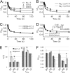
Protons differentially modulate SI onset. A, normalized current amplitude is plotted as a function of prepulse duration and fitted with a double exponential equation (Equation 6) for WT (squares), H880Q (circles), and C373F (triangles) channels at pH 7.4. The asymptote of SI onset at pH 7.4 was significantly different between WT, H880Q, and C373F channels: 43.5 ± 2.2, 48.6 ± 1.2, and 57.9 ± 1.5%, respectively (Table 4). B, pH 6.0 significantly increased τast and τlow but did not affect the relative component of either time constant in WT channels. C, proton modulation of SI recovery was preserved in the H880Q mutant, although the effect on τfast was significantly reduced relative to WT (p < 0.05). D, C373F abolished proton modulation of τfast and τslow; however, pH 6.0 significantly increased the relative component of τslow. The inset in D displays the pulse protocol used. Protons did not significantly affect the asymptote of SI onset in any of the constructs. E and F, bar graphs showing τfast and τslow (E) and relative components of τfast and τslow (F) for SI recovery at pH 7.4 (filled bars) and pH 6.0 (open bars). *, statistically significant at p < 0.05. Error bars, S.E.
FIGURE 6.
FIGURE 6.
Turret mutations abolish proton modulation of UDI. A–C, normalized current is plotted as a function of time and fitted with a double exponential function (Equation 6). Error bars are omitted for image clarity. A, pH 6.0 (gray symbols) significantly increased the τFast from 4.0 ± 0.5 to 7.2 ± 0.7 s and the asymptote of UDI from 60.6 ± 1.1 to 65.3 ± 1.4% in WT channels. B, the C373F mutation removed proton modulation of the UDI asymptote but not τFast. pH 6.0 significantly increased τFast in C373F channels from 2.5 ± 0.2 to 5.0 ± 0.6 s. Additionally, τFast in C373F channels was significantly reduced compared with WT channels (Table 5). C, UDI in H880Q channels was not modulated by protons. D and E, bar graphs depicting τFast (D) and asymptote (E) of UDI of WT, C373F, and H880Q channels at pH 7.4 (filled bars) and pH 6.0 (open bars). The inset of B displays the pulse protocol used. *, statistically significant at p < 0.05. Error bars, S.E.
FIGURE 7.
FIGURE 7.
A, predicted NaV channel secondary structure depicting the relative locations of the C373F (★) and H880Q (♦) mutations. B, sequence alignment of the p-loop segments in bacterial NaChBac, NaVAb, and DI and DII of the mammalian NaV1.2 (accession number Q99250.3) and NaV1.5 (accession number Q14524.2) channels (1). Selectivity residues of NaChBac and NaVAb along with the homologous residues in NaV1.2 and NaV1.5 channels are highlighted in gray. Residues that differ between NaV1.2 and NaV1.5 are encapsulated. P1 and P2 represent the two pore helices isolated in the crystal structure of the NaVAb channel (17, 43).

Similar articles

Cited by

References

    1. Gellens M. E., George A. L., Jr., Chen L. Q., Chahine M., Horn R., Barchi R. L., Kallen R. G. (1992) Primary structure and functional expression of the human cardiac tetrodotoxin-insensitive voltage-dependent sodium channel. Proc. Natl. Acad. Sci. U.S.A. 89, 554–558 - PMC - PubMed
    1. Makita N., Bennett P. B., George A. L. (1996) Molecular determinants of B1 subunit-induced gating modulation in voltage-dependent Na+ channels. J. Neurosci. 16, 7117–7127 - PMC - PubMed
    1. Noda M. (1993) Structure and function of sodium channels. Ann. N.Y. Acad. Sci. 707, 20–37 - PubMed
    1. Jensen M. Ø., Jensen T. R., Kjaer K., Bjørnholm T., Mouritsen O. G., Peters G. H. (2002) Orientation and conformation of a lipase at an interface studied by molecular dynamics simulations. Biophys. J. 98, 111a - PMC - PubMed
    1. Terlau H., Heinemann S. H., Stühmer W., Pusch M., Conti F., Imoto K., Numa S. (1991) Mapping the site of block by tetrodotoxin and saxitoxin of sodium channel II. FEBS Lett. 293, 93–96 - PubMed

Publication types

MeSH terms

LinkOut - more resources