iBet uBet web content aggregator. Adding the entire web to your favor.


Link to original content: https://pubmed.ncbi.nlm.nih.gov/22808031
Phylogenetic interrelationships of ginglymodian fishes (Actinopterygii: Neopterygii) - PubMed Skip to main page content
U.S. flag

An official website of the United States government

Dot gov

The .gov means it’s official.
Federal government websites often end in .gov or .mil. Before sharing sensitive information, make sure you’re on a federal government site.

Https

The site is secure.
The https:// ensures that you are connecting to the official website and that any information you provide is encrypted and transmitted securely.

Access keys NCBI Homepage MyNCBI Homepage Main Content Main Navigation
. 2012;7(7):e39370.
doi: 10.1371/journal.pone.0039370. Epub 2012 Jul 11.

Phylogenetic interrelationships of ginglymodian fishes (Actinopterygii: Neopterygii)

Affiliations

Phylogenetic interrelationships of ginglymodian fishes (Actinopterygii: Neopterygii)

Adriana López-Arbarello. PLoS One. 2012.

Abstract

The Ginglymodi is one of the most common, though poorly understood groups of neopterygians, which includes gars, macrosemiiforms, and "semionotiforms." In particular, the phylogenetic relationships between the widely distributed "semionotiforms," and between them and other ginglymodians have been enigmatic. Here, the phylogenetic relationships between eight of the 11 "semionotiform" genera, five genera of living and fossil gars and three macrosemiid genera, are analysed through cladistic analysis, based on 90 morphological characters and 37 taxa, including 7 out-group taxa. The results of the analysis show that the Ginglymodi includes two main lineages: Lepisosteiformes and †Semionotiformes. The genera †Pliodetes, †Araripelepidotes, †Lepidotes, †Scheenstia, and †Isanichthys are lepisosteiforms, and not semionotiforms, as previously thought, and these taxa extend the stratigraphic range of the lineage leading to gars back up to the Early Jurassic. A monophyletic †Lepidotes is restricted to the Early Jurassic species, whereas the strongly tritoral species previously referred to †Lepidotes are referred to †Scheenstia. Other species previously referred to †Lepidotes represent other genera or new taxa. The macrosemiids are well nested within semionotiforms, together with †Semionotidae, here restricted to †Semionotus, and a new family including †Callipurbeckia n. gen. minor (previously referred to †Lepidotes), †Macrosemimimus, †Tlayuamichin, †Paralepidotus, and †Semiolepis. Due to the numerous taxonomic changes needed according to the phylogenetic analysis, this article also includes formal taxonomic definitions and diagnoses for all generic and higher taxa, which are new or modified. The study of Mesozoic ginglymodians led to confirm Patterson's observation that these fishes show morphological affinities with both halecomorphs and teleosts. Therefore, the compilation of large data sets including the Mesozoic ginglymodians and the re-evaluation of several hypotheses of homology are essential to test the hypotheses of the Halecostomi vs. the Holostei, which is one of the major topics in the evolution of Mesozoic vertebrates and the origin of modern fish faunas.


Conflict of interest statement

Competing Interests: The author has declared that no competing interests exist.

Figures

Figure 1
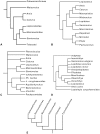
Figure 1. Previous hypothesis of “semionotiform” phylogenetic relationships.
A, Olsen & McCune, 1991 ; B, Gardiner et al. 1996; C, Brito, 1997 ; D, Cavin & Suteethorn, 2006 ; E, Grande, 2010 .
Figure 2
Figure 2. Hypotheses of “semionotiform” phylogenetic relationships of Cavin .
A, Strict consensus tree of 71 most parsimonious trees after the first cladistic analysis based on 31 taxa and 42 informative characters. B, strict consensus tree of 26 most parsimonious trees after the third analysis based on 25 taxa (excluding taxa with 35% or more missing data) and 45 informative characters (three multistate characters were split).
Figure 3
Figure 3. Splint-like quadratojugal. A
, Present and independent in †Macrosemimimus lennieri (BMNHUK P.25180). B, Present and partially fused to the quadrate in †Macrosemius rostratus (BSPG AS-I-770). C, Completely fused to the quadrate or absent in †Thrissops formosus (JME ETT-74). Abbreviations: q, quadrate; qj, quadratojugal. Scale bars point anteriorly.
Figure 4
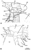
Figure 4. Posterior extension of parietals median to the single pair of laterally placed extrascapular bones.
A, †Macrosemius rostratus (reconstruction based on BSPG AS-I-770; redrawn from [39]). B, †Macrosemimimus fegerti (JME ETT-854). Abbreviations: dpt, dermopteroticum; dsp, dermosphenoticum; ex, extrascapular; fr, frontal; hy, hyomandibula; io, infraorbital bones; op, operculum; pa, parietal; pop, preoperculum; ptt, posttemporal; scl, supracleithrum; suo, suborbital bone.
Figure 5
Figure 5. Variation in the shape of the frontal bones in basal neopterygians. A
, frontal bones distinctly broader posteriorly, but long and narrow anteriorly; redrawn from , fig. 147: restoration of a Callovian species of †Leptolepis (Teleostei) based on isolated bones. B, Broad antorbital portion of frontal; line drawing of the frontals in †Lepidotes laevis MNHN-CRN 61. C, Antorbital portion of frontal tapering gradually; line drawing of the frontals in †Lepidotes minor NHMUK PV P.1118. D, Tubular antorbital portion of frontal; redrawn from , fig. 23: line drawing of the frontals in †Propterus elongatus BSPG 1964-23-145. Drawn to the same size for comparison.
Figure 6
Figure 6. Skull in †Semionotus bergeri.
White arrow indicates the triangular lateral expansion of antorbital portion of frontal in SMF P6108.
Figure 7
Figure 7. Circumborbital bones in neopterygians.
A, †Amia calva redrawn from Grande & Bemis : fig. 16. B, †Propterus elongatus redrawn from : fig. 24. C, †Semionotus elegans redrawn from : fig. 4. D, †Atractosteus spatula redrawn from : fig. 423. Abbreviations: a.io, anterior infraorbital bone; ao, antorbital; dsp, dermosphenoticum; io, infraorbital bone; iop, interoperculum; m.suo, mosaic of suborbital bones; op, operculum; pop, preoperculum; ro, rostral; so, supraorbital; sop, suboperculum; suo, suborbital; t.io, toothed infraorbital bones.
Figure 8
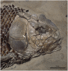
Figure 8. Skull of †Lepidotes gigas Agassiz, 1832 .
Specimen BSPG 1940-I-8 from the area of Holzmaden, Germany.
Figure 9
Figure 9. Details of the skull of †Araripelepidotes temnurus. A
, Anterior portion of the skull in BSPG 1965-I-132 showing the anterior infraorbital. B, Upper and lower jaws in MNHN BCE-336. C (photograph) and D (line drawing), Posterodorsal portion of the skull in AMNH 11833R showing the path of the supraorbital sensory canals. Abbreviations: a.io, anterior infraorbital bone; ao, antorbital; d, dentary; dph, dermosphenoticum; dpt, dermopteroticum; ext, extrascapular; fr, frontal; io, infraorbital bone; l.et, lateral ethmoid; mx, maxilla; n, nasal; op, operculum; ors, orbitosphenoid; pa, parietal; pop, preoperculum; ro, rostral; so, supraorbital; suo, suborbital.
Figure 10
Figure 10. Low number of suborbital bones.
A, Skull of the neotype of †Lepidotes minor (GSM 27975). B, Skull of the holotype of †Tlayuamichin itztli (IGM 6716). Abbreviations: mx, maxilla; op, operculum; sop, suboperculum; suo, suborbital. Arrows indicate the high ascending process of the suboperculum.
Figure 11
Figure 11. Skull in two large tritoral species.
A, †Lepidotes mantelli (NHMUK PV P. 6933). B, †Lepidotes laevis (MNHN-CRN 61).
Figure 12
Figure 12. Opercular bones in †Pliodetes nigeriensis.
A, Holotype specimen MNHN GDF-1275. B, specimen MNHN GDF-1276 showing detached ventral arm of preoperculum. Abbreviations: op, operculum; pop, preoperculum; sop, suboperculum. Scale bars point anteriorly.
Figure 13
Figure 13. Caudal fin in two ginglymodians.
A, †Sangiorgioichthys sui (GMPKU-P-1359). B, †Macrosemimimus fegerti (JME ETT-1351). White arrows indicate the marginal row of scales in the body lobe. The black arrow indicates the scale-like ray.
Figure 14
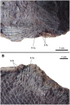
Figure 14. Body scales with strong posterior spine.
A, †Pliodetes nigeriensis (MNHN GDF-1275), B, †Araripelepidotes temnurus (MNHN BCE-335).
Figure 15
Figure 15. Different modes of scale articulation in ginglymodians.
A, †Sangiorgioichthys sui GMPKU-P-1642. B, †Semionotus bergeri (NMC 15128a). C, †Paralepidotus ornatus (BSPG 2003-XXIX-218). D, †Lepidotes minor (NHMUK PV P8047). E, †Lepidotes mantelli (NHMUK PV 2397 and 4916). F, †Araripelepidotes temnurus (MNHN BCE-334). Abbreviations: a.d.pr, anterior dorsal process; a.v.pr, anterior ventral process; d.p, dorsal peg.
Figure 16
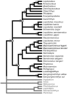
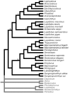
Figure 16. Strict consensus of 69 most parsimonious trees (92 characters, 39 taxa).
Tree length  = 327; consistency index (CI)  = 0.3547; homoplasy index (HI)  = 0.6453; retention index (RI)  = 0.6608; rescaled consistency index (RC)  = 0.2344. Bootstrap and Bremer values are given above and below the branches leading to each node, respectively.
Figure 17
Figure 17. Majority rule consensus of 69 most parsimonious trees (92 characters, 39 taxa).
Tree length  = 327; consistency index (CI)  = 0.3547; homoplasy index (HI)  = 0.6453; retention index (RI)  = 0.6608; rescaled consistency index (RC)  = 0.2344. The numbers above the branches indicate the percentage of MPTs containing the corresponding node.
Figure 18
Figure 18. Results obtained after analyzing Cavin’s data matrices excluding the artificial outgroup and, thus, using Amia and †Leptolepis coryphaeonoides as real out-group taxa. A
, Strict consensus of 26 most parsimonious trees computed with 43 characters and 30 taxa. B, Strict consensus of 24 most parsimonious trees computed with 46 characters and 24 taxa.
Figure 19
Figure 19. †Lepidotes gigas Agassiz, 1832
. BSPG 1940-I-8, SL  = 60.5 cm, from the area of Holzmaden, Germany.
Figure 20
Figure 20. Distinction between: A, †Lepidotes gigas (BSPG 1940-I-8) and B, †Lepidotes elvensis (MNHN JRE-250).
Abbreviations: a.io, anterior infraorbital; ao, antorbital; dph, dermosphenoticum; mx, maxilla; n, nasal; pmx, premaxilla; so, supraorbital; suo, suborbital.
Figure 21
Figure 21. Neotype of †Scheenstia maximus (Wagner, 1863)
. SMF P.2386, SL  = 168 cm, from Solnhofen limestones at Langenaltheim, Bavaria, Germany.
Figure 22
Figure 22. Holotype of †Scheenstia decoratus (Wagner, 1863)
. BSPG AS-VI-3, estimated SL  =  c. 43 cm.
Figure 23
Figure 23. Lectotype of †Callipurbeckia minor (Agassiz, 1833)
. GSM 27975, LS  = 23.5 cm, from the Middle Purbeck Beds at Swanage, Dorset (England).
Figure 24
Figure 24. Fringing and basal fulcra in †Callipurbeckia tendaguruensis (Arratia & Schultze 1999) .
A, Pectoral fin in MBf 7040. B, Dorsal fin in MBf 7041. Abbreviations: b.fu, basal fulcra; fr.fu, fringing fulcrum.
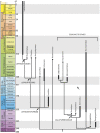
Figure 25
Figure 25. Callibrated phylogenetic hypothesis of ginglymodians interrelationships based on a simplyfied version of the strict consensus tree shown in Figure 17 .
Figure 26
Figure 26. Premaxillary nasal processes in †Pliodetes nigeriensis Wenz, 1999 (MNHN-GDF-1314).
Abbreviations: f.I, foramen for the olfactory nerve; fr, frontal; mx, maxilla; pmx, premaxilla; so, supraorbital. Black arrow points to the suture between the nasal process and the toothed portion of the left premaxilla. Scale bar points anteriorly.

Similar articles

Cited by

References

    1. Regan CT. 24. The Skeleton of Lepidosteus, with remarks on the origin and evolution of the lower Neopterygian Fishes. Proc Zool Soc London. 1923;1923:445–461.
    1. Brough J. The Triassic fishes of Besano, Lombardy. London: The Trustees of the British Museum. pp 117+ VII pl. 1939.
    1. Danil’chenko PG. Obruchev DV, editor. Superorder Holostei. 1964. pp. 378–395. editor. Osnovy Paleontologii. Moskva: Akad. Nauk SSSR. (in Russian).
    1. McAllister DE. The evolution of branchiostegals. Bull Natl Mus Canada. 1968;221:1–239.
    1. Gardiner BG. A revision of certain actinopterygian and coelacanth fishes, chiefly from the Lower Lias. Bulletin of the British Museum (Natural History), Geology. 1960;4:239–384.

Publication types