iBet uBet web content aggregator. Adding the entire web to your favor.


Link to original content: https://pubmed.ncbi.nlm.nih.gov/22623779
Progressive loss of memory T cell potential and commitment to exhaustion during chronic viral infection - PubMed Skip to main page content
U.S. flag

An official website of the United States government

Dot gov

The .gov means it’s official.
Federal government websites often end in .gov or .mil. Before sharing sensitive information, make sure you’re on a federal government site.

Https

The site is secure.
The https:// ensures that you are connecting to the official website and that any information you provide is encrypted and transmitted securely.

Access keys NCBI Homepage MyNCBI Homepage Main Content Main Navigation
. 2012 Aug;86(15):8161-70.
doi: 10.1128/JVI.00889-12. Epub 2012 May 23.

Progressive loss of memory T cell potential and commitment to exhaustion during chronic viral infection

Affiliations

Progressive loss of memory T cell potential and commitment to exhaustion during chronic viral infection

Jill M Angelosanto et al. J Virol. 2012 Aug.

Abstract

T cell exhaustion and loss of memory potential occur during many chronic viral infections and cancer. We investigated when during chronic viral infection virus-specific CD8 T cells lose the potential to form memory. Virus-specific CD8 T cells from established chronic infection were unable to become memory CD8 T cells if removed from infection. However, at earlier stages of chronic infection, these virus-specific CD8 T cells retained the potential to partially or fully revert to a memory differentiation program after transfer to infection-free mice. Conversely, effector CD8 T cells primed during acute infection were not protected from exhaustion if transferred to a chronic infection. We also tested whether memory and exhausted CD8 T cells arose from different subpopulations of effector CD8 T cells and found that only the KLRG1(lo) memory precursor subset gave rise to exhausted CD8 T cells. Together, these studies demonstrate that CD8 T cell exhaustion is a progressive developmental process. Early during chronic infection, the fate of virus-specific CD8 T cells remains plastic, while later, exhausted CD8 T cells become fixed in their differentiation state. Moreover, exhausted CD8 T cells arise from the memory precursor and not the terminally differentiated subset of effector CD8 T cells. These studies have implications for our understanding of senescence versus exhaustion and for therapeutic interventions during chronic infection.


Figures
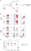
Fig 1
Fig 1
Virus-specific CD8 T cells at day 30 after LCMV clone 13 infection do not persist if adoptively transferred to infection-free recipients. (A) Experimental design. Ly5.1+ CD8 T cells were purified from day 30 LCMV clone 13-infected mice and adoptively transferred to day 30 LCMV Arm-immune or day 30 LCMV clone 13-infected recipient mice. Approximately 2 × 10⁵ DbGP33+ CD8 T cells were adoptively transferred to each recipient mouse. Donor virus-specific CD8 T cells were examined on days 2 and 60 p.t. (B) Gating strategy used to identify donor CD8 T cell population. Representative examples of the day 30 clone 13 → Arm and day 30 clone 13 → clone 13 recipient mice on both day 2 p.t. and day 60 p.t. (C) Absolute numbers of DbGP33+ CD8 T cells in the spleens of recipient mice on days 2 and 60 p.t. normalized to the number of cells on day 2 p.t. Each symbol represents one mouse, with red squares signifying antigen-specific CD8 T cells transferred to clone 13-infected recipient mice and blue circles representing cells transferred to day 30 Arm-infected recipient mice. ns, not significant; **, P < 0.01.
Fig 2
Fig 2
CD8 T cells from day 15 clone 13-infected mice persisted but formed memory poorly in infection-free recipients. (A) Experimental design. Ly5.2+ CD8 T cells were purified from day 15 LCMV clone 13-infected mice and adoptively transferred to day 15 Arm- or day 15 clone 13-infected recipient mice. Approximately 1 × 10⁵ or 1.5 × 10⁵ donor DbGP33+ CD8 T cells were adoptively transferred to each recipient mouse, with equal numbers of adoptively transferred cells transferred to all donor mice for each individual experiment. Donor virus-specific CD8 T cells were examined on days 5, 15, 30, and 60 p.t. in recipient mouse spleens. (B) Representative fluorescence-activated cell sorter plots of CD44hi DbGP33+ day 15 clone 13 → Arm-infected and day 15 clone 13 → clone 13-infected recipient mice on day 60 p.t. The cells are gated on live CD8+ Ly5.2+ T cells. (C) Mean fluorescence of CD127 and PD-1. The gray histogram in each plot is gated on CD8+ CD44lo naïve T cells. (D) Frequency of PD-1hi and CD127+ CD8+ DbGP33+ T cells on day 60 p.t. (E) Mean fluorescence of CD127 and PD-1 on days 5, 15, and 30 p.t. (F) Percentage of IFN-γ, IL-2, TNF, and IFN-γ plus TNF produced by CD8 T cells following restimulation ex vivo with the GP33 peptide on day 30 p.t. Light blue, endogenous (Endo) Arm response; dark blue, clone 13 → Arm transfer; red, clone 13 → clone 13 transfer; pink, endogenous clone 13 response.
Fig 3
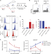
Fig 3
Virus-specific CD8 T cells isolated from LCMV clone 13-infected donor mice on day 8 p.i. can form memory in infection-free recipient mice. (A) Experimental design. Ly5.2+ CD8 T cells were purified from day 8 clone 13-infected mice and adoptively transferred to day 8 Arm- or day 8 clone 13-infected recipient mice. In each experiment, the same number of CD8 T cells was transferred to each recipient mouse, with between 2 × 10⁵ and 3 × 10⁵ donor DbGP33+ CD8 T cells transferred to each recipient per experiment. Donor virus-specific CD8 T cells were examined on days 5, 15, 30, and 60 p.t. in recipient mouse spleens. (B) Representative fluorescence-activated cell sorter plots of CD44hi DbGP33+ day 8 clone 13 → Arm-infected and day 8 clone 13 → clone 13-infected recipient mice on day 30 p.t. The cells are gated on live CD8+ Ly5.2+ T cells. (C) Mean fluorescence of CD127 and PD-1. The gray histogram in each plot is gated on CD8+ CD44lo naïve T cells. (D) Frequency of PD-1hi and CD127+ CD8+ DbGP33+ T cells on day 60 p.t. (E) Mean fluorescence of CD127 and PD-1 on days 5, 15, and 30 p.t. Light blue, endogenous Arm response; dark blue, clone 13 → Arm transfer; red, clone 13 → clone 13 transfer; pink, endogenous clone 13 response.
Fig 4
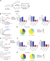
Fig 4
Optimal priming does not prevent exhaustion of effector CD8 T cells transferred to a chronic infection. (A) Experimental design. Ly5.1+ CD8 T cells were purified from either day 8 or day 15 Arm-infected mice and adoptively transferred (day 8, ∼5 × 10⁵ DbGP33+ CD8 T cells/recipient; day 15, ∼1 × 10⁵ DbGP33+ CD8 T cells/recipient) to infection-matched clone 13- or Arm-infected recipient mice. Donor virus-specific CD8 T cells were examined on days 5, 15, 30, and 60 p.t. in recipient spleens. (B to D) Day 8 Arm → clone 13. (B) Percentage of Ly5.1+ DbGP33+ CD8 T cells expressing CD127 or high levels of PD-1 in the blood; (C) mean fluorescence of IFN-γ, IL-2, and TNF expressed by CD8 T cells restimulated ex vivo with the GP33 peptide on day 60 p.t.; (D) polyfunctionality of day 8 Arm → Arm and day 8 Arm → clone 13. Colors represent the number of cytokines (1 to 5) coproduced per cell. (E to G) Day 15 Arm → clone 13. (E) Percentage of Ly5.1+ DbGP33+ CD8 T cells expressing CD127 or high levels of PD-1 in the blood; (F) mean fluorescence of IFN-γ, IL-2, and TNF expressed by CD8 T cells restimulated ex vivo with the GP33 peptide on day 60 p.t.; (G) polyfunctionality of day 15 Arm → Arm and day 15 Arm → clone 13 displayed as in panel D. MFI, mean fluorescence intensity; MIP1α, macrophage inflammatory protein-1α.
Fig 5
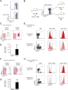
Fig 5
Exhausted virus-specific CD8 T cells arise from the KLRG1lo memory precursor population of effector cells. (A) KLRG1 expression on day 8 p.i. with Arm and clone 13. (B) Experimental design. Ly5.2+ CD44hi DbGP33+ CD8 T cells from either day 8 Arm- or day 8 clone 13-infected donor mice were sorted into KLRG1hi and KLRG1lo populations. Equal numbers of virus-specific KLRG1hi- or KLRG1lo-sorted populations were adoptively transferred into day 8 clone 13-infected recipient mice and analyzed at 1 to 2 months p.t. Approximately 2.9 × 10⁵ KLRG1hi or KLRG1lo DbGP33+ CD8 T cells were transferred to each recipient in the Arm → clone 13 experiment, and 1.4 × 10⁵ cells were transferred in the clone 13 → clone 13 control experiment. (C to E) KLRG1hi and KLRG1lo CD8+ DbGP33+ T cells from day 8 Arm-infected mice adoptively transferred into day 8 clone 13-infected recipients. (C) Representative plots of the endogenous (Ly5.2-negative [Ly5.2]) and KLRG1-sorted (Ly5.2+) donor populations at 2 months p.t.; (D) absolute number of each transferred population on day 60 p.t.; (E) representative plots from KLRG1lo recipient mice on day 60 p.t. The mean fluorescence of CD127 and PD-1 is shown in red or pink, with CD8+ CD44lo naïve T cells shown in the gray histograms. (F and G) KLRG1hi or KLRG1lo CD8+ DbGP33+ T cells from day 8 clone 13-infected mice adoptively transferred into day 8 clone 13-infected recipients. (F) Representative plots of the endogenous (Ly5.2) and KLRG1-sorted (Ly5.2+) donor populations at 2 months p.t.; (G) absolute number of each transferred population on day 60 p.t. (H) Representative plots from KLRG1lo recipient mice on day 60 p.t. The mean fluorescence of CD127 and PD-1 is shown in red or pink, with CD8+ CD44lo naïve T cells shown in the gray histograms.

Similar articles

Cited by

References

    1. Ahmed R, Jamieson BD, Porter DD. 1987. Immune therapy of a persistent and disseminated viral infection. J. Virol. 61:3920–3929 - PMC - PubMed
    1. Ahmed R, Salmi A, Butler LD, Chiller JM, Oldstone MB. 1984. Selection of genetic variants of lymphocytic choriomeningitis virus in spleens of persistently infected mice. Role in suppression of cytotoxic T lymphocyte response and viral persistence. J. Exp. Med. 160:521–540 - PMC - PubMed
    1. Akbar AN, Henson SM. 2011. Are senescence and exhaustion intertwined or unrelated processes that compromise immunity? Nat. Rev. Immunol. 11:289–295 - PubMed
    1. Alter G, et al. 2003. Longitudinal assessment of changes in HIV-specific effector activity in HIV-infected patients starting highly active antiretroviral therapy in primary infection. J. Immunol. 171:477–488 - PubMed
    1. Appay V, Almeida JR, Sauce D, Autran B, Papagno L. 2007. Accelerated immune senescence and HIV-1 infection. Exp. Gerontol. 42:432–437 - PubMed

Publication types

MeSH terms