Human herpesvirus-6 entry into the central nervous system through the olfactory pathway
- PMID: 21825120
- PMCID: PMC3158203
- DOI: 10.1073/pnas.1105143108
Human herpesvirus-6 entry into the central nervous system through the olfactory pathway
Abstract
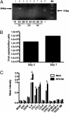
Viruses have been implicated in the development of neurodegenerative diseases, such as Alzheimer's, Parkinson's, and multiple sclerosis. Human herpesvirus-6 (HHV-6) is a neurotropic virus that has been associated with a wide variety of neurologic disorders, including encephalitis, mesial temporal lobe epilepsy, and multiple sclerosis. Currently, the route of HHV-6 entry into the CNS is unknown. Using autopsy specimens, we found that the frequency of HHV-6 DNA in the olfactory bulb/tract region was among the highest in the brain regions examined. Given this finding, we investigated whether HHV-6 may infect the CNS via the olfactory pathway. HHV-6 DNA was detected in a total of 52 of 126 (41.3%) nasal mucous samples, showing the nasal cavity is a reservoir for HHV-6. Furthermore, specialized olfactory-ensheathing glial cells located in the nasal cavity were demonstrated to support HHV-6 replication in vitro. Collectively, these results support HHV-6 utilization of the olfactory pathway as a route of entry into the CNS.
Conflict of interest statement
The authors declare no conflict of interest.
Figures


Similar articles
-
Detection of human herpesvirus-6 in mesial temporal lobe epilepsy surgical brain resections.Neurology. 2003 Nov 25;61(10):1405-11. doi: 10.1212/01.wnl.0000094357.10782.f9. Neurology. 2003. PMID: 14638964 Free PMC article.
-
The role of herpesvirus 6A and 6B in multiple sclerosis and epilepsy.Scand J Immunol. 2020 Dec;92(6):e12984. doi: 10.1111/sji.12984. Epub 2020 Oct 23. Scand J Immunol. 2020. PMID: 33037649 Free PMC article. Review.
-
Glycoprotein gE-negative pseudorabies virus has a reduced capability to infect second- and third-order neurons of the olfactory and trigeminal routes in the porcine central nervous system.J Gen Virol. 1994 Nov;75 ( Pt 11):3095-106. doi: 10.1099/0022-1317-75-11-3095. J Gen Virol. 1994. PMID: 7964619
-
Association of human herpesvirus-6B with mesial temporal lobe epilepsy.PLoS Med. 2007 May;4(5):e180. doi: 10.1371/journal.pmed.0040180. PLoS Med. 2007. PMID: 17535102 Free PMC article.
-
Human herpesvirus-6 entry into host cells.Future Microbiol. 2010 Jul;5(7):1015-23. doi: 10.2217/fmb.10.61. Future Microbiol. 2010. PMID: 20632802 Review.
Cited by
-
Association between human herpesviruses infections and childhood neurodevelopmental disorders: insights from two-sample mendelian randomization analyses and systematic review with meta-analysis.Ital J Pediatr. 2024 Nov 20;50(1):248. doi: 10.1186/s13052-024-01820-9. Ital J Pediatr. 2024. PMID: 39568007 Free PMC article.
-
The Development of Epilepsy Following CNS Viral Infections: Mechanisms.Curr Neurol Neurosci Rep. 2024 Nov 16;25(1):2. doi: 10.1007/s11910-024-01393-4. Curr Neurol Neurosci Rep. 2024. PMID: 39549124 Review.
-
Symptomatic central nervous system tuberculosis and human herpesvirus-6 coinfection with associated hydrocephalus managed with endoscopic third ventriculostomy: A case report and review of human herpesvirus-6 neuropathology.Surg Neurol Int. 2024 Aug 16;15:287. doi: 10.25259/SNI_355_2024. eCollection 2024. Surg Neurol Int. 2024. PMID: 39246759 Free PMC article.
-
Herpesvirus Infection of Endothelial Cells as a Systemic Pathological Axis in Myalgic Encephalomyelitis/Chronic Fatigue Syndrome.Viruses. 2024 Apr 8;16(4):572. doi: 10.3390/v16040572. Viruses. 2024. PMID: 38675914 Free PMC article. Review.
-
Identification of a strong genetic risk factor for major depressive disorder in the human virome.iScience. 2024 Feb 10;27(3):109203. doi: 10.1016/j.isci.2024.109203. eCollection 2024 Mar 15. iScience. 2024. PMID: 38414857 Free PMC article.
References
-
- Mori I, Nishiyama Y, Yokochi T, Kimura Y. Olfactory transmission of neurotropic viruses. J Neurovirol. 2005;11(2):129–137. - PubMed
-
- Bach LM. Regional physiology of the central nervous system. Prog Neurol Psychiatry. 1963;18:46–106. - PubMed
-
- Chowdhury SI, Lee BJ, Onderci M, Weiss ML, Mosier D. Neurovirulence of glycoprotein C(gC)-deleted bovine herpesvirus type-5 (BHV-5) and BHV-5 expressing BHV-1 gC in a rabbit seizure model. J Neurovirol. 2000;6:284–295. - PubMed
-
- Liedtke W, Opalka B, Zimmermann CW, Lignitz E. Age distribution of latent herpes simplex virus 1 and varicella-zoster virus genome in human nervous tissue. J Neurol Sci. 1993;116(1):6–11. - PubMed
MeSH terms
Substances
LinkOut - more resources
Full Text Sources
Other Literature Sources


