iBet uBet web content aggregator. Adding the entire web to your favor.


Link to original content: https://pubmed.ncbi.nlm.nih.gov/21349843
The phosphoinositide kinase PIKfyve is vital in early embryonic development: preimplantation lethality of PIKfyve-/- embryos but normality of PIKfyve+/- mice - PubMed Skip to main page content
U.S. flag

An official website of the United States government

Dot gov

The .gov means it’s official.
Federal government websites often end in .gov or .mil. Before sharing sensitive information, make sure you’re on a federal government site.

Https

The site is secure.
The https:// ensures that you are connecting to the official website and that any information you provide is encrypted and transmitted securely.

Access keys NCBI Homepage MyNCBI Homepage Main Content Main Navigation
. 2011 Apr 15;286(15):13404-13.
doi: 10.1074/jbc.M111.222364. Epub 2011 Feb 24.

The phosphoinositide kinase PIKfyve is vital in early embryonic development: preimplantation lethality of PIKfyve-/- embryos but normality of PIKfyve+/- mice

Affiliations

The phosphoinositide kinase PIKfyve is vital in early embryonic development: preimplantation lethality of PIKfyve-/- embryos but normality of PIKfyve+/- mice

Ognian C Ikonomov et al. J Biol Chem. .

Abstract

Gene mutations in the phosphoinositide-metabolizing enzymes are linked to various human diseases. In mammals, PIKfyve synthesizes PtdIns(3,5)P(2) and PtdIns5P lipids that regulate endosomal trafficking and responses to extracellular stimuli. The consequence of pikfyve gene ablation in mammals is unknown. To clarify the importance of PIKfyve and PIKfyve lipid products, in this study, we have characterized the first mouse model with global deletion of the pikfyve gene using the Cre-loxP approach. We report that nearly all PIKfyve(KO/KO) mutant embryos died before the 32-64-cell stage. Cultured fibroblasts derived from PIKfyve(flox/flox) embryos and rendered pikfyve-null by Cre recombinase expression displayed severely reduced DNA synthesis, consistent with impaired cell division causing early embryo lethality. The heterozygous PIKfyve(WT/KO) mice were born at the expected Mendelian ratio and developed into adulthood. PIKfyve(WT/KO) mice were ostensibly normal by several other in vivo, ex vivo, and in vitro criteria despite the fact that their levels of the PIKfyve protein and in vitro enzymatic activity in cells and tissues were 50-55% lower than those of wild-type mice. Consistently, steady-state levels of the PIKfyve products PtdIns(3,5)P(2) and PtdIns5P selectively decreased, but this reduction (35-40%) was 10-15% less than that expected based on PIKfyve protein reduction. The nonlinear decrease of the PIKfyve protein versus PIKfyve lipid products, the potential mechanism(s) discussed herein, may explain how one functional allele in PIKfyve(WT/KO) mice is able to support the demands for PtdIns(3,5)P(2)/PtdIns5P synthesis during life. Our data also shed light on the known human disorder linked to PIKFYVE mutations.


Figures
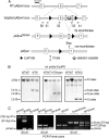
FIGURE 1.
FIGURE 1.
Targeted disruption of mouse pikfyve gene. A, schematic representation of the strategy used for the conditional knock-out of the pikfyve gene by excision of exon 6. Shown is the locus between exons 5 and 7. Indicated are the sites of the PCR-specific primers S0–S3 and A1 used for genotyping. B, representative Southern blots of genomic DNA derived from mice with the indicated genotypes and digested with EcoRV. Membranes were probed with a genomic probe called “en probe” (indicated). C, representative genotyping of mice and embryos by PCR using the specific primer pairs indicated in A. The expected fragment sizes for the wild-type, KO, and floxΔneo alleles are indicated in parentheses. templ, template.
FIGURE 2.
FIGURE 2.
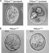
Preimplantation PIKfyveKO/KO embryos are abnormal. A and B, embryos from the PIKfyveWT/KO intercrosses were collected at the E3.5 stage, observed by phase-contrast, and then genotyped by nested PCR. Arrows in A denote the presence of aberrant vacuoles in the PIKfyveKO/KO embryos. Also apparent is the decreased cell number in both the inner cell mass and trophectoderm.
FIGURE 3.
FIGURE 3.
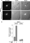
Arrested DNA synthesis in flox/flox MEFs rendered pikfyve-null by expression of Cre recombinase. A and B, cultured MEFs (100-mm dish) isolated from E12.5 PIKfyveWT/WT and PIKfyveflox/flox embryos were transfected by electroporation with the Cre recombinase-GFP cDNA and then seeded on coverslips. A, 72 h post-transfection, cells were observed by a 40× Hoffman objective. Shown are typical phase-contrast images of the defective vacuolar phenotype due to Cre recombinase-induced pikfyve gene deletion (panels a–c) seen in 96 ± 4% (mean ± S.E.) of Cre-transfected flox/flox MEFs. Panels d–f illustrate the normal morphology that was observed in nearly all of the Cre-transfected wild-type cells. Predominant Cre expression is seen in the nucleus (panels c and d). B, 36 h post-transfection, cells were serum-deprived for 36 h and then stimulated for 18 h in the presence or absence of FBS (20%). BrdU was included during the last 6 h of incubation, and its cell incorporation was detected by indirect immunofluorescence with an anti-BrdU and Alexa Fluor 568-conjugated anti-mouse IgG as primary and secondary antibodies, respectively. GFP/BrdU fluorescence-positive MEFs from three experiments were counted, and the mitogen response is presented as a percentage of the total GFP-Cre-transfected cells. In Cre-expressing flox/flox MEFs, serum-induced BrdU incorporation was practically not apparent in contrast to the Cre-expressing wild-type cells, which readily incorporated BrdU under these conditions. Error bars, ± S.E.
FIGURE 4.
FIGURE 4.
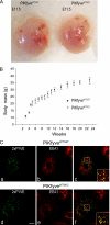
PIKfyveWT/KO embryos, mice, and endosomal system are similar to wild type. A, embryos from the PIKfyveWT/KO intercrosses were collected at E11.5, photographed, and genotyped. B, body weights of male PIKfyveWT/KO and wild-type littermates born from PIKfyveWT/KO intercrosses fed on mouse chow (11% fat); error bars, ± S.E. C, E12.5 embryos from the PIKfyveWT/KO intercrosses were dissected. MEFs were then isolated and cultured. MEFs collected from a confluent 100-mm dish were transfected by electroporation with the pEGFP-2xFYVE cDNA for staining the PtdIns3P-enriched endosomes. Cells were then reseeded and processed 24 h post-transfection by confocal microscopy with anti-EEA1 antibodies. The morphologic appearance of the endosomal system and the extent of colocalization (insets in the merged images c and f) between EEA1- (panels b and e) and 2xFYVE-associated fluorescence (panels a and d) in PIKfyveWT/WT and PIKfyveWT/KO MEFs are similar. Bar, 10 μm.
FIGURE 5.
FIGURE 5.
PIKfyve protein and in vitro activity are reduced by 2-fold in PIKfyveWT/KO. A, RIPA+ lysates derived from cultured MEFs (E12.5) or from the indicated tissues dissected from PIKfyveWT/KO mice and PIKfyveWT/WT littermates were clarified by centrifugation. Equal amounts of tissue protein (200 μg) and MEF lysates (150 μg) from each genotype were examined by immunoblotting with anti-PIKfyve antibodies. Blots were reprobed with anti-tubulin or anti-actin for loading. Shown are chemiluminescence detections from representative experiments with two embryos/animals for each condition of two to three independent determinations. *, an unspecific band in the muscle samples. WB, Western blot. B and C, fresh RIPA+ lysates derived from MEFs or brain tissues dissected from PIKfyveWT/KO and PIKfyveWT/WT embryos (E12.5) and mice, respectively, underwent immunoprecipitation (IP) with anti-PIKfyve antibodies. Washed immunoprecipitates were subjected to in vitro lipid kinase activity assay. Shown are a representative autoradiogram of a TLC plate with separated radiolabeled lipids (MEFs) (B) and quantitation of the lipid kinase activities in MEFs and brain by radioactive counting of the scraped silica corresponding to PtdIns5P and PtdIns(3,5)P2 (C). Presented are the combined decreases of both lipids as the extent of their individual decrease in PIKfyveWT/KO versus wild type was identical. D, MEFs prepared from PIKfyveWT/KO (panels b and d) and PIKfyveWT/WT embryos (E12.5) (panels a and c) were seeded on coverslips. Cells were then treated for 2 h with YM201636 (0–160 nm) and observed live by the 40× objective of the Hoffman Modulation Contrast System for progression of a vacuolation phenotype. The phase-contrast images illustrate that only the PIKfyveWT/KO MEFs exhibit the typical defective vacuolar phenotype under these conditions.
FIGURE 6.
FIGURE 6.
PIKfyve lipid products are decreased to lesser degree than PIKfyve protein in PIKfyveWT/KO MEFs. MEFs isolated from E12.5 PIKfyveWT/KO and PIKfyveWT/WT embryos and seeded on 35-mm dishes were subjected to inositol and glucose starvation (22 h). Cells were then labeled with myo-[2-3H]inositol (26 h) as described under “Experimental Procedures.” Lipids were extracted, deacylated, and fractionated by HPLC. Presented are the radioactivity from the HPLC profiles of a representative experiment with both genotypes (A) and the quantitation (B) from four independent experiments calculated as a percentage of the total phosphoinositide radioactivity and presented as mean ± S.E. The total phosphoinositide (PI) radioactivity was obtained by summing the counts within the elution times corresponding to the indicated [3H]GroPIns peaks. The changes in PtdIns(3,5)P2, PtdIns5P, and PtdIns3P in PIKfyveWT/KO versus PIKfyveWT/WT were statistically significant (*) with p = 0.0014, p = 0.0076, and p = 0.0488, respectively.
FIGURE 7.
FIGURE 7.
Decreased PAS complexes in PIKfyveWT/KO MEFs. Cultured MEFs isolated from E12.5 PIKfyveWT/KO and PIKfyveWT/WT embryos were solubilized in RIPA+. Clarified lysates underwent immunoprecipitation (IP) with anti-PIKfyve antibodies. After washes in the same buffer, immunoprecipitates were analyzed by SDS-PAGE and immunoblotting. Blots, cut horizontally above the 110-kDa standard, were probed with anti-PIKfyve (top part), anti-Sac3, and anti-ArPIKfyve antibodies (bottom part) with a stripping step in between. The top and bottom parts were probed with anti-IRAP (insulin-responsive aminopeptidase, ∼160 kDa) and anti-α-tubulin antibodies (∼55 kDa), respectively, for equal loading. Shown are chemiluminescence detections of a representative experiment of three independent determinations.

Similar articles

Cited by

References

    1. Balla T. (2006) J. Endocrinol. 188, 135–153 - PubMed
    1. Di Paolo G., De Camilli P. (2006) Nature 443, 651–657 - PubMed
    1. Vicinanza M., D'Angelo G., Di Campli A., De Matteis M. A. (2008) Cell. Mol. Life Sci. 65, 2833–2841 - PMC - PubMed
    1. Ooms L. M., Horan K. A., Rahman P., Seaton G., Gurung R., Kethesparan D. S., Mitchell C. A. (2009) Biochem. J. 419, 29–49 - PubMed
    1. Majerus P. W., York J. D. (2009) J. Lipid Res. 50, (suppl.) S249–S254 - PMC - PubMed

Publication types

MeSH terms