iBet uBet web content aggregator. Adding the entire web to your favor.


Link to original content: https://pubmed.ncbi.nlm.nih.gov/21192805
The normal development of Platynereis dumerilii (Nereididae, Annelida) - PubMed Skip to main page content
U.S. flag

An official website of the United States government

Dot gov

The .gov means it’s official.
Federal government websites often end in .gov or .mil. Before sharing sensitive information, make sure you’re on a federal government site.

Https

The site is secure.
The https:// ensures that you are connecting to the official website and that any information you provide is encrypted and transmitted securely.

Access keys NCBI Homepage MyNCBI Homepage Main Content Main Navigation
. 2010 Dec 30:7:31.
doi: 10.1186/1742-9994-7-31.

The normal development of Platynereis dumerilii (Nereididae, Annelida)

Affiliations

The normal development of Platynereis dumerilii (Nereididae, Annelida)

Antje Hl Fischer et al. Front Zool. .

Abstract

Background: The polychaete annelid Platynereis dumerilii is an emerging model organism for the study of molecular developmental processes, evolution, neurobiology and marine biology. Annelids belong to the Lophotrochozoa, the so far understudied third major branch of bilaterian animals besides deuterostomes and ecdysozoans. P. dumerilii has proven highly relevant to explore ancient bilaterian conditions via comparison to the deuterostomes, because it has accumulated less evolutionary change than conventional ecdysozoan models. Previous staging was mainly referring to hours post fertilization but did not allow matching stages between studies performed at (even slightly) different temperatures. To overcome this, and to provide a first comprehensive description of P. dumerilii normal development, a temperature-independent staging system is needed.

Results: Platynereis dumerilii normal development is subdivided into 16 stages, starting with the zygote and ending with the death of the mature worms after delivering their gametes. The stages described can be easily identified by conventional light microscopy or even by dissecting scope. Developmental landmarks such as the beginning of phototaxis, the visibility of the stomodeal opening and of the chaetae, the first occurrence of the ciliary bands, the formation of the parapodia, the extension of antennae and cirri, the onset of feeding and other characteristics are used to define different developmental stages. The morphology of all larval stages as well as of juveniles and adults is documented by light microscopy. We also provide an overview of important steps in the development of the nervous system and of the musculature, using fluorescent labeling techniques and confocal laser-scanning microscopy. Timing of each developmental stage refers to hours post fertilization at 18 ± 0.1°C. For comparison, we determined the pace of development of larvae raised at 14°C, 16°C, 20°C, 25°C, 28°C and 30°C. A staging ontology representing the comprehensive list of developmental stages of P. dumerilii is available online.

Conclusions: Our atlas of Platynereis dumerilii normal development represents an important resource for the growing Platynereis community and can also be applied to other nereidid annelids.


Figures
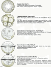
Figure 1
Figure 1
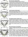
Schemes of developmental stages of P. dumerilii: zygote, cleaving embryo, stereoblastula - stereogastrula, protrochophore and early trochophore. Left: The scheme indicates the key characteristics of each developmental stage. Right: Next to the scheme a brief summary of the key features for the stage is given. The time points indicated with a star mark the end of each stage and are excluded from this stage. Bold: The name of the stage and in brackets the beginning and end of each stage is given. Italic: The key characteristics, which can be used to determine the beginning of each stage. Normal: Additional features of each stage are given, including some landmarks, which can be seen in the developing nervous system and musculature. From top to bottom: zygote (apical view), cleaving embryo (apical view), stereoblastula - stereogastrula (ventral view, apical up), protrochophore (ventral view, apical up) and early trochophore (ventral view, apical up). Abbreviations see abbreviations list.
Figure 2
Figure 2
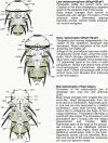
Schemes of developmental stages of P. dumerilii: mid-trochophore, late trochophore, early metatrochophore and mid-metatrochophore. Left: The scheme indicates the key characteristics of each developmental stage. Right: Next to the scheme a brief summary of the key features for the stage is given. The time points indicated with a star mark the end of each stage and are excluded from this stage. Bold: The name of the stage and in brackets the beginning and end of each stage is given. Italic: The key characteristics, which can be used to determine the beginning of each stage. Normal: Additional features of each stage are given, including some landmarks, which can be seen in the developing nervous system and musculature. From top to bottom: mid-trochophore, late trochophore, early metatrochophore, mid-metatrochophore. All schemes shown as ventral view, anterior up. Abbreviations see abbreviations list.
Figure 3
Figure 3
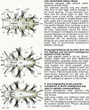
Schemes of developmental stages of P. dumerilii: late metatrochophore, early nectochaete and mid-nectochaete. Left: The scheme indicates the key characteristics of each developmental stage. Right: Next to the scheme a brief summary of the key features for the stage is given. The time points indicated with a star mark the end of each stage and are excluded from this stage. Bold: The name of the stage and in brackets the beginning and end of each stage is given. Italic: The key characteristics, which can be used to determine the beginning of each stage. Normal: Additional features of each stage are given, including some landmarks, which can be seen in the developing nervous system and musculature. From top to bottom: late metatrochophore, early nectochaete and mid-nectochaete. All schemes shown as ventral view, anterior left. Abbreviations see abbreviations list.
Figure 4
Figure 4
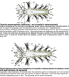
Schemes of developmental stages of P. dumerilii: late nectochaete, three-segmented errant juvenile and 4- and 5-segmented errant juveniles. Left: The scheme indicates the key characteristics of each developmental stage. Right: Next to the scheme a brief summary of the key features for the stage is given. Bold: The name of the stage and in brackets the beginning and end of each stage is given. Italic: The key characteristics, which can be used to determine the beginning of each stage. Normal: Additional features of each stage are given. From top to bottom: late nectochaete, three-segmented errant juvenile, Four- and five-segmented errant juvenile. All schemes shown as ventral view, anterior left. Abbreviations see abbreviations list.
Figure 5
Figure 5
Schemes of developmental stages of P. dumerilii: cephalic metamorphosis and small atokous worm. Top: The scheme indicates the key characteristics of each developmental stage. Below each scheme: A brief summary of the key features for the stage is given. Bold: The name of the stage and in brackets the beginning and end of each stage is given. Italic: The key characteristics, which can be used to determine the beginning of each stage. Normal: Additional features of each stage are given. From top to bottom: cephalic metamorphosis, small atokous worm. All schemes shown as ventral view, anterior left. Abbreviations see abbreviations list.
Figure 6
Figure 6
Schemes of developmental stages of P. dumerilii: Sexual metamorphosis: male and female/heteronereis. Top: The scheme indicates the key characteristics of the mature male and female heteronereis. Bottom: A brief summary of the key features for this stage is given. Bold: The name of the stage and in brackets the beginning and end of the stage is given. Italic: The key characteristics, which can be used to determine the beginning of each stage. Normal: Additional features of each stage are given. Schemes are shown as dorsal view, anterior left (schemes are modified from: [37]). Abbreviations see abbreviations list.
Figure 7
Figure 7
Ventral nerve cord and muscle development of P. dumerilii, 24-30 hpf, ventral view, anterior up. The age of the larvae in each row is given in the lower left corner of the first picture of each row. The displayed staining is indicated at the bottom of each column. A, G, M, S: Two axons/axon bundles, which grow from posterior to anterior, connect the posterior serotonergic cell to the prototroch ring nerve. They form the connectives (con) of the ventral nerve cord. B: The first serotonergic cell of the ventral nerve cord is located at the posterior end of the larva (white star). C, D, I, J, O, P, U, V: The dorsal longitudinal muscles are visible form 28 hpf onwards. E, K, Q, W: The stomodeal field (sf) is visible on the ventral side. F, L, R, X: The telotroch (tt) is visible at the posterior end of the larva from around 26 hpf onwards. CLSM microscopy, maximum projection, Imaris surpass mode. Scale bar in all images 20 μm. Further abbreviations see abbreviations list.
Figure 8
Figure 8
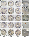
Series of conventional light microscopy images of P. dumerilii, 24-36 hpf. For each time point an overview (left) and the larval eye at higher magnification (right) is shown. All images except O and P are apical view of larvae, dorsal side up. O: Ventro-posterior view of the larva at 36 hpf, apical side left. P: Anterior-ventral view of a larva at 36 hpf, apical side left. A, C, E, G, I, K, M, O: In all larva the 4 macromeres (mm) in the center of the larva is visible. Each macromere contains one lipid droplet (ld). The two ventral lipid droplets are smaller than the dorsal ones. Around the prototroch additional pigment (pi) is visible in some larvae (I, K, M, O). B, D, F, H, J, L, N, P: The pigmentation of the larval eyes (le) becomes more intense over time. Scale bar in all images 20 μm. Further abbreviations see abbreviations list. In this and all other figure plates, fresh samples were used at every time point. Each specimen was collected from 18°C ± 0.1°C source immediately before imaging.
Figure 9
Figure 9
Brain and muscle development of P. dumerilii, 24-30 hpf, apical view, dorsal side up. The age of the larvae in each row is given in the lower left corner of the first picture of each row. The displayed staining is indicated at the bottom of each column. A, F, K, P: The dorsal root of the circumesophageal connectives (drcc) is visible at 24 hpf as well as the developing prototroch ring nerve (ptrn). From 26 hpf onwards the ventral branch of the circumesophageal connectives (vrcc) and the unpaired dorsal axon (ua) are visible. The dorsal and ventral roots of the circumesophageal connectives connect the cerebral commissures (cc) in the brain. The prototroch ring nerve becomes more pronounced. B, G, L: The first serotonergic cell (white star) is located in the apical organ just below the apical tuft (apt). Q: At 30 hpf a second serotonergic cell appears (white arrow). C, D, H, I, M, N, R, S: From 28 hpf onwards the dorsal longitudinal muscles (dlm) become visible. E, J, O, T: The amount of cells in the brain is increasing. CLSM microscopy, maximum projection, Imaris surpass mode. Scale bar in all images 20 μm. Further abbreviations see abbreviations list.
Figure 10
Figure 10
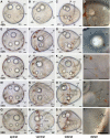
Series of conventional light microscopy images of P. dumerilii, 38-46 hpf. The age of the larvae in each row is given in the lower left corner of the first picture of each row. To give an overview on the morphological changes throughout development, for each time point an apical view of larvae, dorsal side up (first column: A, E, I, M, Q), ventral view, anterior side left (second column: B, F, J, N, R), lateral view, anterior side left and dorsal side up (third column: C, G, K, O, S) and several details (fourth column: D, H, L, P, T) are shown. H: From 40 hpf onwards the chaetae (ch) (white arrow heads) can be seen inside the trunk. L, T: The chaetae (white arrow heads) grow out from the chaetal sacs and increase in length (chs). L: The chaetal sacs are marked with a white stippled line on the right side of the larva. E, N: The stomodeal opening is visible. Note that the intensity of the pigmentation pattern may vary substantially around the prototroch and posterior end among trochophore larvae. F, J, N: Individuals with a high amount of pigment, whereas the individuals in B and R show almost no pigment. Scale bar in all images 20 μm. Further abbreviations see abbreviations list.
Figure 11
Figure 11
Ventral nerve cord and muscle development of P. dumerilii, 32-38 hpf, ventral view, anterior up. The age of the larvae in each row is given in the lower left corner of the first picture of each row. The displayed staining is indicated at the bottom of each column. A, G, M, S: The connectives (con) show an increasing staining intensity (compare the tubulin staining at the different time points). S: The first commissure (1com) of the ventral nerve cord is formed. B, H, N, T: Only one serotonergic cell is visible in the ventral nerve cord (white star). C, D, G, D, I, J, O, P, U, V: The dorsal longitudinal muscles (dlm) increase in length and the ventral longitudinal muscles (vlm) become clearly visible by phalloidin staining at 32 hpf. E, F, K, L, Q, R, W, X: The stomodeal field (sf) gets located more anteriorly between 32 hpf and 38 hpf and cells start to form a circle in the stomodeal field. CLSM microscopy, maximum projection, Imaris surpass mode. Scale bar in all images 20 μm. Further abbreviations see abbreviations list.
Figure 12
Figure 12
Brain and muscle development of P. dumerilii, 32-38 hpf, apical view, dorsal side up. The age of the larvae in each row is given in the lower left corner of the first picture of each row. The displayed staining is indicated at the bottom of each column. A, F, K, P: The dorsal and ventral branch of the circumesophageal connectives (drcc and vrcc) become thicker and the prototroch ring nerve (ptrn) is well visible. The dorsal and ventral roots of the circumesophageal connectives connect the cerebral commissures (cc). B, G, L, Q: Additionally to the first and second serotonergic cell (white star and white arrow respectively) two more serotonergic cells develop laterally in the brain of the larvae (white arrows). C, D, H, I, M, N, R, S: The dorsal longitudinal muscles (dlm) become more pronounced. The ventral longitudinal muscles (vlm) become clearly visible around 32 hpf. E, J, O, T: The amount of cells on the ventral side of the episphere is increasing. CLSM microscopy, maximum projection, Imaris surpass mode. Scale bar in all images 20 μm. Further abbreviations see abbreviations list.
Figure 13
Figure 13
Ventral nerve cord and muscle development of P. dumerilii, 40-46 hpf, ventral view, anterior up. The age of the larvae in each row is given in the lower left corner of the first picture of each row. The displayed staining is indicated at the bottom of each column. A, G, M, S: The second commissure (2com) of the ventral nerve cord is formed at 42 hpf. B, H, N, T: In addition to the unpaired serotonergic cell at the posterior end of the larva (white star), a pair of serotonergic neurons becomes visible at the first commissure (white arrow head). C, D, I, J, O, P, U, V: The dorsal and ventral longitudinal muscles (dlm and vlm) increase rapidly in length. E, F, K, L, Q, R, W, X: The stomodeal rosette (str) is formed and gets into a more anterior position. CLSM microscopy, maximum projection, Imaris surpass mode. Scale bar in all images 20 μm. Further abbreviations see abbreviations list.
Figure 14
Figure 14
Brain and muscle development of P. dumerilii, 40-46 hpf, apical view, dorsal side up. The age of the larvae in each row is given in the lower left corner of the first picture of each row. The displayed staining is indicated at the bottom of each column. A, F, K, P: The dorsal and ventral branch of the circumesophageal connectives (drcc and vrcc), the cerebral commissures (cc) and the dorsal unpaired axon (ua) are well visible, the prototroch ring nerve (ptrn) is well developed. B, G, L, Q: Four serotonergic cells are visible in the brain (the central one marked with a white star, the more dorsal ones marked with white arrows). G, D, H, I, M, N, R, S: The dorsal longitudinal muscles (dlm) and ventral longitudinal muscles (vlm) are intensely stained. E, J, O, T: The stomodeum (sto) becomes visible in the episphere from around 42 hpf onwards. CLSM microscopy, maximum projection, Imaris surpass mode. Scale bar in all images 20 μm. Further abbreviations see abbreviations list.
Figure 15
Figure 15
Ventral nerve cord and muscle development of P. dumerilii, 48-54 hpf, ventral view, anterior up. The age of the larvae in each row is given in the lower left corner of the first picture of each row. The displayed staining is indicated at the bottom of each column. A, G, M, S: The third commissure (3com) of the ventral nerve cord is formed at 48 hpf and more commissural axons are visible in the ventral nerve cord (e.g. compare A, G, M and S). B, H, N, T: In addition to the unpaired serotonergic cell at the posterior end of the larva (white star) and the first pair of serotonergic neurons at the first commissure. A pair of serotonergic cells near the second commissure and a second pair at the first commissure become visible (white arrow heads). E, K, Q, W: The stomodeal rosette (str) is well visible and contains more cells over time. C, D, I, J, O, P, U, V: Parapodial muscles (ppm) and oblique muscles (om) become from 48 hpf onwards. F, L, R, X: The first paratroch (pat1) is visible at the posterior boarder of the second chaetigerous segment from around 48 hpf onwards. CLSM microscopy, maximum projection, Imaris surpass mode. Scale bar in all images 20 μm. Further abbreviations see abbreviations list.
Figure 16
Figure 16
Brain and muscle development of P. dumerilii, 48-54 hpf, apical view, dorsal side up. The age of the larvae in each row is given in the lower left corner of the first picture of each row. The displayed staining is indicated at the bottom of each column. A, F, K, P: The dorsal and ventral branches of the circumesophageal connectives (drcc and vrcc) approach each other. The distance between the prototroch ring nerve (ptrn) and the prototroch (pt) begins to increase and the prototroch ring nerve approaches the cerebral commissures (cc) (e.g. compare A, F, K and P). B, G, L, Q: Four serotonergic cells are visible in the brain (the central one marked with a white star, the more dorsal ones marked with white arrows). C, D, H, I, M, N, R, S: The dorsal longitudinal muscles (dlm) and ventral longitudinal muscles (vlm) are intensely stained. E, J, O, T: The stomodeum is well visible in the episphere. CLSM microscopy, maximum projection, Imaris surpass mode. Scale bar in all images 20 μm. Further abbreviations see abbreviations list.
Figure 17
Figure 17
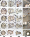
Series of conventional light microscopy images of P. dumerilii, 48-56 hpf. The arrangement of the images is similar to figure 10. H: At 50 hpf the adult eye pigment (ae) appears very faintly. L, M, O, P, Q, S: Adult eye pigment becomes well visible from 52 hpf onwards. J: The chaetae (ch) break through the body wall at 52 hpf. F, J, N, R: Chaetae increase rapidly in length. T: Additionally to the first paratroch (with arrows) the second paratroch (black arrows) develop at the posterior boarder of the first segment. Scale bar in all images 20 μm. Further abbreviations see abbreviations list.
Figure 18
Figure 18
Series of conventional light microscopy images of P. dumerilii, 58-66 hpf. The arrangement of the images is similar to figure 10. B, F, J, N, R: The chaetae (ch) rapidly increase in length and the chaetae of the third segment reach the posterior end of the larva, which marks the beginning of the late metatrochophore stage. F, J, N, R: Thee pairs of parapodia (pp) start forming laterally at the trunk. D, H, L, P: The amount of pigment in the adult eyes (ae) increases. T: The metatroch (black arrows) forms posterior to the prototroch at 66 hpf. Scale bar in all images 20 μm. Further abbreviations see abbreviations list.
Figure 19
Figure 19
Ventral nerve cord and muscle development of P. dumerilii, 56-62 hpf, ventral view, anterior up. The age of the larvae in each row is given in the lower left corner of the first picture of each row. The displayed staining is indicated at the bottom of each column. A, G, M, S: An increasing amount of commissural axons becomes visible along the ventral nerve cord (compare A, G, M, S). B, H, N, T: A pair of serotonergic cells becomes visible in the third segment (white arrow heads), additionally to the unpaired serotonergic cell (white star) and other serotonergic cells in the first and second segment (white arrow heads). C, D, I, J, O, P, U, V: Parapodial muscles (ppm) and oblique muscles (om) increase rapidly in size. The unpaired medial ventral longitudinal muscle (mvlm) becomes visible posterior to the stomodeum and elongates posteriorly (compare C, I, O, U). E, K, Q, W: The stomodeal rosette (str) is well visible. Three pairs of parapodia (pp) become visible from around 60 hpf onwards. F, L, R, X: The second paratroch (pat2) becomes visible at the posterior boarder of the first chaetigerous segment from 56 hpf onwards. CLSM microscopy, maximum projection, Imaris surpass mode. Scale bar in all images 20 μm. Further abbreviations see abbreviations list.
Figure 20
Figure 20
Brain and muscle development of P. dumerilii, 56-62 hpf, apical view, dorsal side up. The age of the larvae in each row is given in the lower left corner of the first picture of each row. The displayed staining is indicated at the bottom of each column. A, F, K, P: The dorsal and ventral branches of the circumesophageal connectives (drcc and vrcc) continue to approach each other. The distance between the prototroch ring nerve (ptrn) and the prototroch (pt) increases (compare A, F, K, P). B, G, L, Q: Four serotonergic cells are visible in the brain (the central one marked with a white star, the more dorsal ones marked with white arrows). C, D, H, I, M, N, R, S: The dorsal longitudinal muscles (dlm) and ventral longitudinal muscles (vlm) are intensely stained. E, J, O, T: Dorsally, the first ciliated tufts of the akrotroch (act) develop (compare J and O). The akrotroch is a row of ciliated cells, which goes laterally from the prototroch dorso-medially onto the head. CLSM microscopy, maximum projection, Imaris surpass mode. Scale bar in all images 20 μm. Further abbreviations see abbreviations list.
Figure 21
Figure 21
Ventral nerve cord and muscle development of P. dumerilii, 64 - 70 hpf, ventral view, anterior up. The age of the larvae in each row is given in the lower left corner of the first picture of each row. The displayed staining is indicated at the bottom of each column. A, G, M, S: The connectives (con) and commissures (com) are intensely stained. The connectives have a band-like shape. B, H, N, T: Additional serotonergic cells develop in the ventral nerve cord (white arrow heads), in addition to the unpaired serotonergic cell (white star). C, D, I, J, O, P, U, V: Parapodial muscles (ppm) and oblique muscles (om) as well as the unpaired medial ventral longitudinal muscle (mvlm) are well visible. E, K, Q, W: The metatroch (mt) becomes visible posterior to the prototroch (pt) around 66 hpf. F, L, R, X: The parapodia (pp) grow in size. CLSM microscopy, maximum projection, Imaris surpass mode. Scale bar in all images 20 μm. Further abbreviations see abbreviations list.
Figure 22
Figure 22
Brain and muscle development of P. dumerilii, 64-70 hpf, apical view, dorsal side up. The age of the larvae in each row is given in the lower left corner of the first picture of each row. The displayed staining is indicated at the bottom of each column. A, F, K, P: The dorsal and ventral branches of the circumesophageal connectives (drcc and vrcc) continue to approach each other. The distance between the prototroch ring nerve (ptrn) and the prototroch (pt) increases further (e.g. compare A, F, K and P). Therefore, the overall appearance of the brain is more compact. B, G, L, Q: As it was the case during earlier time points, four serotonergic cells are visible in the brain (the central one marked with a white star, the more dorsal ones marked with white arrows). C, D, H, I, M, N, R, S: Musculature around the stomodeum (stm) forms an arch, which branch off the ventral longitudinal muscles (vlm). E, J, O, T: Additional ciliated cells become visible in the akrotroch to form a line on either side of the dorsal head. CLSM microscopy, maximum projection, Imaris surpass mode. Scale bar in all images 20 μm. Further abbreviations see abbreviations list.
Figure 23
Figure 23
Ventral nerve cord and muscle development of P. dumerilii, 72 hpf - 5 days post fertilization, ventral view, anterior up. The age of the larvae in each row is given in the lower left corner of the first picture of each row. The displayed staining is indicated at the bottom of each column. A, G, M: The ventral nerve cord is well visible. The connectives become broader. Therefore, the commissures appear shorter than in previous stages. The segmental nerves (sn) become well visible. Antennal nerves (an) and palpi nerves (pln) are visible. B, H, N: More serotonergic cells develop in the ventral nerve cord (white arrow heads). The unpaired serotonergic cell, which was marked by a white star in figure 7, 10, 11, 13, 15, 18, 21, 19, 26 and 28 is not visible anymore. C, D, I, J, O, P: Parapodial muscles (ppm) and oblique muscles (om) as well as the unpaired medial ventral longitudinal muscle (mvlm) are well visible and a complex set of muscles develop in the head region, e.g. the antennal muscles (antm). E, K, Q: The dorsal branch of the anterior cirri (adc) begins to elongate. CLSM microscopy, maximum projection, Imaris surpass mode. Scale bar in all images 20 μm. Further abbreviations see abbreviations list.
Figure 24
Figure 24
Brain and muscle development of P. dumerilii, 72 hpf - 5 days post fertilization, apical view, dorsal side up. The age of the larvae in each row is given in the lower left corner of the first picture of each row. The displayed staining is indicated at the bottom of each column. A, E, F, J, K, O: The dorsal and ventral branches of the circumesophageal connectives (drcc and vrcc) continue to approach each other. Finally only a small gap remains between the dorsal and ventral branch of the circumesophageal connectives at 5 days post fertilization (L). The prototroch ring nerve (ptrn) and the dorsal unpaired axon (ua) cannot be distinguished anymore from 5 days post fertilization onwards with the methods used in this study (L). B, G, L: New serotonergic cells become visible in the dorsal part of the brain at 4 and 5 days post fertilization (white arrow heads), additionally to those serotonergic cells that were present during previous stages (the central one marked with a white star, the more dorsal ones marked with white arrows). C, D, I, M, N: A complex pattern of muscles develops inside the head. CLSM microscopy, maximum projection, Imaris surpass mode. Scale bar in all images 20 μm. Further abbreviations see abbreviations list.
Figure 25
Figure 25
Series of conventional light microscopy images of P. dumerilii, 68-72 hpf. The arrangement of images is similar to figure 10. B, F, J: The trunk of the larvae elongates rapidly. D, H: The pigmentation in the adult eyes (ae) increases. C, G, K: The lipid droplets (ld) start to move posteriorly. B, F, J: The stomodeum (sto) appears slit-like due its invagination. Scale bar in all images 20 μm. Further abbreviations see abbreviations list.
Figure 26
Figure 26
Series of conventional light microscopy images of P. dumerilii, 75 hpf (mid nectochaete) - late nectochaete. A-D: larvae at 75 hpf. A: Ventral view of larvae, anterior side left. B: Detail of the developing antennae (ant). C: Detail of the developing anterior dorsal cirrus (adc). D: Detail of the developing anal cirri and the forming midgut (mg) and proctodeum (proc). E, J and O: Dorsal view of larvae, anterior side left. The age is indicated in the lower left corner of each picture. The antennae (ant), anterior dorsal cirri (adc) and anal cirri (ac) increase in length. The lipid droplets (ld) get absorbed. F-I, K-N and P-S: Several details of larvae at 4 days, 5 days and 6 days respectively. Compare A, F, K and P: The adult eyes (ae) increase in size compared to previous stages. H, M, R: The jaw (j) is increasing in size and the primary tooth (1to) becomes well visible. I, N, S: The proctodeum (proc) forms a connection to the midgut (mg) at around 5 days. O: Food particles (fp) are clearly visible in the midgut (mg). The palpi (pl) are visible as circular structures. G, L, Q: The larval eyes are remaining. Scale bar in all images 20 μm. Further abbreviations see abbreviations list.
Figure 27
Figure 27
Series of conventional light microscopy images of P. dumerilii, 3-segmented errant juvenile - small atokous worm with 7 chaetigerous segments. The stage of the juvenile in each row is given in the lower left corner of the first picture of each row. In order to give an overview on the morphological changes throughout development, for each time point a dorsal view, anterior side left (first column: A, D, G, J, M), a close-up of the head (second column: B, E, H, K, N) and several details (third column: C, F, I, L, O) are shown. B: Anterior view of juvenile, dorsal side up. E, H, K and N: Dorsal view of juvenile, anterior side left. Compare A, E, H, K and N: Note the transformation of the most anterior pair of parapodia into the dorsal pair of posterior cirri (pdc). While the number of segments is indicated with CS4, CS5 respectively before cephalic metamorphosis, the numbers are indicated with CS4', CS5' after cephalic metamorphosis. Please note, that CS5 and CS5' are not the same segment. A more detailed description of the cephalic metamorphosis is given in the text. C and I: Stomodeum at a higher magnification, the jaws (j) increase in size and additional teeth (th) form. F and L: All secondary segments are formed by the posterior growth zone (gz). Scale bar in A, D, G, J, M: 100 μm, in all other images 20 μm. Further abbreviations see abbreviations list.
Figure 28
Figure 28
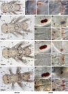
Series of conventional light microscopy images of P. dumerilii, small atokous worm and heteronereis. All images: Dorsal view, anterior side left. A: Overview, small atokous worm with 48 chaetigerous segments. B: Head of the same specimen as shown in A, at a higher magnification. The dorsal and ventral branch of the anterior and posterior cirri are visible, as well as the palpi (pl), antennae (ant) and the well developed jaws (j). C: Posterior end of the same specimen at higher magnification, posterior to the last chaetigerous segment is the growth zone (gz) located where new segments are formed. D: Overview, male and female may be similarly sized. Female (♀), top, appears yellowish, due to the oocytes in the coelomic cavity. Male (♂) bottom, appears white due to spermatozoans in the anterior part and red due to dense accessory blood vessels in the posterior part. The parapodia (pp) in the posterior body part are flattened with paddle-like chaetae. E: Detail of the male and female body at a higher magnification. Note the numerous visible oocytes (ooc) inside the female's body and the different shape of the parapodia (pp) in the anterior and posterior body part. F: Anterior part of a mature male. Note the enlarged adult eyes (ae) in comparison to B. The dorsal cirri of the parapodia (dcpp) in segment three to nine are clearly club-shaped. D: Anterior part of a mature female, note also here the enlarged adult eyes (ae) and the oocytes inside the coelomic cavity and the parapodia. The dorsal cirri of the parapodia (dcpp) in segment three to seven are slightly club-shaped. Scale bar in A: 500 μm. Scale bar in B and C: 100 μm. Scale bar in D, E, F and G: 2 mm. Further abbreviations see abbreviations list.
Figure 29
Figure 29
Influence of temperature on developmental speed in P. dumerilii. Diagram, which shows the developmental stage reach 12 h, 24 h, 36 h, 48 h, 60 h and 72 h post fertilization, growing at 14°C, 16°C, 18°C, 20°C, 25°C, 28°C and 30°C. Sampling was performed every 12 h for 72 h. The error bars indicate the range within the stage, at which the embryos or larvae were found. Within this range it was not possible to determine the stage more precisely. The gradient in the chart background indicates the decreasing synchrony from the late nectochaete stage onwards. The transversal lines indicate the end of one stage and the beginning of the following one, The duration of the late nectochaete stage and the beginning of the following stage vary, indicated by the bold blurred line.

Similar articles

Cited by

References

    1. Hutchinson TH, Jha AN, Dixon DR. The Polychaete Platynereis dumerilii (Audouin and Milne-Edwards): A New Species for Assessing the Hazadrous Potential of Chemicals in the Marine Environment. Ecotoxicology and Environmental Safety. 1995;31:271–281. doi: 10.1006/eesa.1995.1074. - DOI - PubMed
    1. Jékely G, Colombelli J, Hausen H, Guy K, Stelzer E, Nédélec F, Arendt D. Mechanism of phototaxis in marine zooplankton. Nature. 2008;456:395–400. - PubMed
    1. Tessmar-Raible K, Arendt D. Emerging systems: between vertebrates and arthropods, the Lophotrochozoa. Current Opinion in Genetics & Development. 2003;13:331–340. - PubMed
    1. Hardege JD. Nereidid polychaetes as model organisms for marine chemical ecology. Hydrobiologia. 1999;402:145–161. doi: 10.1023/A:1003740509104. - DOI
    1. Dorresteijn AWC. Quantitative analysis of cellular differentiation during early embryogenesis of Platynereis dumerilii. Roux's Archives of Developmental Biology. 1990;199:14–30. - PubMed

LinkOut - more resources