Natural history of perihematomal edema after intracerebral hemorrhage measured by serial magnetic resonance imaging
- PMID: 21164136
- PMCID: PMC3074599
- DOI: 10.1161/STROKEAHA.110.590646
Natural history of perihematomal edema after intracerebral hemorrhage measured by serial magnetic resonance imaging
Abstract
Background and purpose: knowledge on the natural history and clinical impact of perihematomal edema (PHE) associated with intracerebral hemorrhage is limited. We aimed to define the time course, predictors, and clinical significance of PHE measured by serial magnetic resonance imaging.
Methods: patients with primary supratentorial intracerebral hemorrhage ≥ 5 cm(3) underwent serial MRIs at prespecified intervals during the first month. Hematoma (H(v)) and PHE (E(v)) volumes were measured on fluid-attenuated inversion recovery images. Relative PHE was defined as E(v)/H(v). Neurologic assessments were performed at admission and with each MRI. Barthel Index, modified Rankin scale, and extended Glasgow Outcome scale scores were assigned at 3 months.
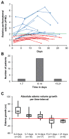
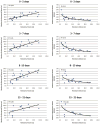
Results: twenty-seven patients with 88 MRIs were prospectively included. Median H(v) and E(v) on the first MRI were 39 and 46 cm(3), respectively. Median peak absolute E(v) was 88 cm(3). Larger hematomas produced a larger absolute E(v) (r(2)=0.6) and a smaller relative PHE (r(2)=0.7). Edema volume growth was fastest in the first 2 days but continued until 12 ± 3 days. In multivariate analysis, a higher admission hematocrit was associated with a greater delay in peak PHE (P=0.06). Higher admission partial thromboplastin time was associated with higher peak rPHE (P=0.02). Edema volume growth was correlated with a decline in neurologic status at 48 hours (81 vs 43 cm(3), P=0.03) but not with 3-month functional outcome.
Conclusions: PHE volume measured by MRI increases most rapidly in the first 2 days after symptom onset and peaks toward the end of the second week. The timing and magnitude of PHE volume are associated with hematologic factors. Its clinical significance deserves further study.
Figures


Similar articles
-
Peak Edema Extension Distance: An Edema Measure Independent from Hematoma Volume Associated with Functional Outcome in Intracerebral Hemorrhage.Neurocrit Care. 2024 Jun;40(3):1089-1098. doi: 10.1007/s12028-023-01886-z. Epub 2023 Nov 29. Neurocrit Care. 2024. PMID: 38030878 Free PMC article.
-
Fingolimod for the treatment of intracerebral hemorrhage: a 2-arm proof-of-concept study.JAMA Neurol. 2014 Sep;71(9):1092-101. doi: 10.1001/jamaneurol.2014.1065. JAMA Neurol. 2014. PMID: 25003359 Clinical Trial.
-
Temporal pattern of cytotoxic edema in the perihematomal region after intracerebral hemorrhage: a serial magnetic resonance imaging study.Stroke. 2013 Apr;44(4):1144-6. doi: 10.1161/STROKEAHA.111.000056. Epub 2013 Feb 7. Stroke. 2013. PMID: 23391767
-
Perihematomal Edema After Intracerebral Hemorrhage: An Update on Pathogenesis, Risk Factors, and Therapeutic Advances.Front Immunol. 2021 Oct 19;12:740632. doi: 10.3389/fimmu.2021.740632. eCollection 2021. Front Immunol. 2021. PMID: 34737745 Free PMC article. Review.
-
Acute evacuation of 54 intracerebral hematomas (aICH) during the microsurgical clipping of a ruptured middle cerebral artery bifurcation aneurysm-illustration of the individual clinical courses and outcomes with a serial brain CT/MRI panel until 12 months.Acta Neurochir (Wien). 2024 Jan 17;166(1):17. doi: 10.1007/s00701-024-05902-9. Acta Neurochir (Wien). 2024. PMID: 38231317 Free PMC article. Review.
Cited by
-
Hemorrhagic stroke in children.J Cent Nerv Syst Dis. 2024 Nov 1;16:11795735241289913. doi: 10.1177/11795735241289913. eCollection 2024. J Cent Nerv Syst Dis. 2024. PMID: 39493255 Free PMC article. Review.
-
Based on hematoma and perihematomal tissue NCCT imaging radiomics predicts early clinical outcome of conservatively treated spontaneous cerebral hemorrhage.Sci Rep. 2024 Aug 9;14(1):18546. doi: 10.1038/s41598-024-69249-y. Sci Rep. 2024. PMID: 39122887 Free PMC article.
-
Chemical Composition and Neuroprotective Properties of Indonesian Stingless Bee (Geniotrigona thoracica) Propolis Extract in an In-Vivo Model of Intracerebral Hemorrhage (ICH).Nutrients. 2024 Jun 14;16(12):1880. doi: 10.3390/nu16121880. Nutrients. 2024. PMID: 38931235 Free PMC article.
-
Early Celecoxib Use in Spontaneous Intracerebral Hemorrhage is Associated with Reduced Mortality.Neurocrit Care. 2024 Dec;41(3):788-797. doi: 10.1007/s12028-024-01996-2. Epub 2024 May 15. Neurocrit Care. 2024. PMID: 38750392
-
The relationship between perihematomal edema and hematoma expansion in acute spontaneous intracerebral hemorrhage: an exploratory radiomics analysis study.Front Neurosci. 2024 Apr 30;18:1394795. doi: 10.3389/fnins.2024.1394795. eCollection 2024. Front Neurosci. 2024. PMID: 38745941 Free PMC article.
References
-
- Xi G, Keep RF, Hoff JT. Mechanisms of brain injury after intracerebral haemorrhage. Lancet Neurol. 2006;5:53–63. - PubMed
-
- Mayer SA, Sacco RL, Shi T, Mohr JP. Neurologic deterioration in noncomatose patients with supratentorial intracerebral hemorrhage. Neurology. 1994;44:1379–1384. - PubMed
-
- Ladurner G, Sager WD, Iliff LD, Lechner H. A correlation of clinical findings and CT in ischaemic cerebrovascular disease. Eur Neurol. 1979;18:281–288. - PubMed
Publication types
MeSH terms
Grants and funding
LinkOut - more resources
Full Text Sources
Medical


