iBet uBet web content aggregator. Adding the entire web to your favor.


Link to original content: https://pubmed.ncbi.nlm.nih.gov/21040849
Extracellular calcium controls background current and neuronal excitability via an UNC79-UNC80-NALCN cation channel complex - PubMed Skip to main page content
U.S. flag

An official website of the United States government

Dot gov

The .gov means it’s official.
Federal government websites often end in .gov or .mil. Before sharing sensitive information, make sure you’re on a federal government site.

Https

The site is secure.
The https:// ensures that you are connecting to the official website and that any information you provide is encrypted and transmitted securely.

Access keys NCBI Homepage MyNCBI Homepage Main Content Main Navigation
. 2010 Nov 4;68(3):488-99.
doi: 10.1016/j.neuron.2010.09.014.

Extracellular calcium controls background current and neuronal excitability via an UNC79-UNC80-NALCN cation channel complex

Affiliations

Extracellular calcium controls background current and neuronal excitability via an UNC79-UNC80-NALCN cation channel complex

Boxun Lu et al. Neuron. .

Abstract

In contrast to its extensively studied intracellular roles, the molecular mechanisms by which extracellular Ca(2+) regulates the basal excitability of neurons are unclear. One mechanism is believed to be through Ca(2+)'s interaction with the negative charges on the cell membrane (the charge screening effect). Here we show that, in cultured hippocampal neurons, lowering [Ca(2+)](e) activates a NALCN channel-dependent Na(+)-leak current (I(L-Na)). The coupling between [Ca(2+)](e) and NALCN requires a Ca(2+)-sensing G protein-coupled receptor, an activation of G-proteins, an UNC80 protein that bridges NALCN to a large novel protein UNC79 in the same complex, and the last amino acid of NALCN's intracellular tail. In neurons from nalcn and unc79 knockout mice, I(L-Na) is insensitive to changes in [Ca(2+)](e), and reducing [Ca(2+)](e) fails to elicit the excitatory effects seen in the wild-type. Therefore, extracellular Ca(2+) influences neuronal excitability through the UNC79-UNC80-NALCN complex in a G protein-dependent fashion.


Figures

Figure 1
Figure 1. Dependence of the Excitatory Action of Low [Ca2+]e on NALCN
A hyperpolarizing holding current (IHold, -45 pA in the neuron in panel A; -30 pA in panel B; -33.7 ± 11.0 pA (wild-type) and -14.8 ± 5.0 pA (NALCN-/-) for panel C) was injected to bring each neuron's steady membrane potential to -80 mV in 1.2 mM Ca2+-containing bath. Pulses (10 s, as illustrated in lower right in panel B) of additional depolarizing currents with increasing amplitudes (+10 to +40 pA; “+injection” in the X-axes) superimposed on the holding currents (IHold) were injected every 50 s. (A, B) Examples of current-clamp recordings from a wild-type (A) and a NALCN mutant neuron (B) in baths containing 1.2 mM (upper traces) or 0.5 mM (lower traces) Ca2+. Firing frequencies of the neurons during the 10 s depolarizing pulses are plotted in the right panels. (C) Statistics of firing frequencies from wild-type (left) and NALCN-/- neurons (right). Error bars, mean and s.e.m. See also Figure S1.
Figure 2
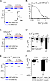
Figure 2. Control of Resting Na+ Leak Current by Extracellular Ca2+ in Cultured Hippocampal Neurons
(A) Representative holding currents at –68 mV in a wild-type (+/+) neuron. Na+-leak current is presented as ΔIL-Na (indicated by the double arrow), defined as the difference between holding currents in 140 mM (solid bar) and 14 mM (open bar) Na+-containing baths. A 0.25 sec recording is shown for each condition. ΔIL-Na increased when [Ca2+]e was switched from 2 mM (indicated by the hatched bar labeled 2 Ca) to 0.1 mM (0.1 Ca). (B) ΔIL-Na in wild-type neurons measured at various [Ca2+]e normalized to that measured with 2 mM [Ca2+]e (n ≥ 5). (C) Similar to (A), but from a NALCN-/- neuron. (D) Comparison of ΔIL-Na between wild-type (+/+) and NALCN-/- neurons at a range of [Ca2+]e, as indicated. The number of cells for each condition is indicated in parentheses. (E) Representative ΔIL-Na restored by NALCN cDNA transfection into the NALCN-/- neurons. (F) Summary of ΔIL-Na generated by NALCN or mock (empty vector) transfection in 2 mM and 0.1 mM [Ca2+]e (mean ± SEM). See also Figure S2.
Figure 3
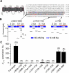
Figure 3. Dependence of Low [Ca2+]e-activated Current (ILCA) on the Carboxy-terminal Residues of NALCN
(A) Schematic illustration of the location of the NALCN C-terminal mutants. The C-terminal sequences (from the rat isoform, accession # NP_705894) are shown (right). Non-conserved (red) and conserved (blue) amino acid substitutions in the chicken isoform (accession # XP_416967) are highlighted. Shadowed sequences indicate sequence non-essential to ILCA (deleted in Δ1623-1699). (B) Representative ΔIL-Na recordings from SH-SY5Y cells transfected with NALCN deletion mutants Δ1623-1699 (with amino acids 1623-1699 deleted), Δ1724-1732 and Δ1738. (C) Summary of potentiation of ΔIL-Na by lowering [Ca2+]e from 2 mM to 0.1 mM, defined as percentage of increase ([ΔIL-Na in 0.1 mM Ca2+ - ΔIL-Na in 2 mM Ca2+]/ΔIL-Na in 2 mM Ca2+). Data from transfected NALCN-/- and SH-SY5Y cells were pooled. Measurements from the 5 full-length NALCN-transfected neurons used in Figure 2F were also included for comparison. See Figure S3 for the averaged sizes of ΔIL-Na in 2 mM and 0.1 mM Ca2+ -containing baths.
Figure 4
Figure 4. Synergism between Low [Ca2+]e and Substance P
(A) Representative recordings of ILCA in the presence and absence of substance P (1 μM), from a wild-type neuron cultured on pre-plated glial cells (left), or a NALCN-/- neuron cultured under the same conditions (right). (B) Average ILCA of wild-type (+/+), NALCN-/-, and full-length (NALCN-/-; NALCN) or carboxy-terminal truncated (Δ1638-1738) NALCN cDNA-transfected NALCN-/- neurons in the presence or absence of SP. For the wild-type or transfected neurons, only cells with greater than 20 pA SP-activated current (ISP, measured under 2 mM [Ca2+]e as illustrated by an arrow in panel A) were selected for analysis. NALCN-/- neurons had no detectable ISP.
Figure 5
Figure 5. UNC79 Forms a Complex with NALCN via Its Interaction with UNC80 in the Brain and Influences UNC80 Protein Level
(A) Association of NALCN, UNC79, and UNC80 in the brain. Total mouse brain protein was immunoprecipitated (IP) with the indicated antibodies and blotted (IB) with anti-NALCN (left) or anti-UNC80 (right) antibodies. Anti-HA (α-Ctrl1) and anti-CATSPER1 (α-Ctrl2) were used as control antibodies for specificity. (B) Association of UNC79 with the NALCN complex via its interaction with UNC80. Lysates from HEK293T cells transfected with the indicated combinations of plasmids were immunoprecipitated and blotted with indicated antibodies (lanes 1–2 and lanes 3–5 are from two separate gels). A FLAG-tagged transmembrane protein, CATSPERβ (FLAG-Ctrl3), was used as a control. (C) Western blot using total brain protein from wild-type (WT) and UNC79 knockout (KO), showing the recognition of native UNC79 protein by the anti-UNC79 antibody. Cell lysates from HEK293T cells transfected with UNC79 cDNA or empty vector (mock) were loaded for molecular weight comparison and assessment of antibody specificity. (D) Western blot showing absence of detectable UNC80 protein in the UNC79 knockout brain. (E) Western blot with anti-NALCN showing NALCN protein in the UNC79 KO (left two lanes). Immunoprecipitating with anti-UNC79 or anti-UNC80 failed to precipitate NALCN in the KO because of the absence of UNC79 and UNC80 in the mutant. See also Figure S7 for a model of the interaction among NALCN, UNC79 and UNC80.
Figure 6
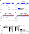
Figure 6. The Na+-leak Current Is Insensitive to [Ca2+]e in UNC79 Mutant Neurons, but the Sensitivity Can Be Rescued with UNC80
(A–C) Representative ΔIL-Na in wild-type (A, +/+), UNC79 knockout (A, UNC79-/-), UNC79-/- transfected with UNC79 cDNA (B), or UNC79-/- transfected with UNC80 (C) neurons in baths containing 2 mM or 0.1 mM Ca2+. (D) Summary of ΔIL-Na recorded with 2 mM [Ca2+]e (left group) and 0.1 mM [Ca2+]e (middle group), and the difference between the two (ILCA, right group). Neurons cultured from littermates under identical conditions were used for comparison between the wild-type and mutant. The number of cells for each condition is indicated in parentheses.
Figure 7
Figure 7. Dependence of the Excitatory Action of Low [Ca2+]e on UNC79
Hyperpolarizing holding currents (IHold, -60 pA in the neuron in panel A; -100 pA in panel B; -64.0 + 18.8 pA (wild-type), -83.7 + 19.2 pA (UNC79-/-) and -32.7 + 17.4 pA (NALCN-/-) for panel C) was injected to bring each neuron's steady membrane potential to -80 mV in 1.2 mM Ca2+-containing bath. Pulses (10 s, as illustrated in lower right in panel B) of additional depolarizing currents with increasing amplitudes (+10 to +60 pA) were injected every 50 s. (A, B) Examples of current-clamp recordings from a wild-type (A) and an UNC79 mutant neuron (B) in baths containing 1.2 mM (upper traces) or 0.1 mM (lower traces) Ca2+. Firing frequencies of the neurons during the 10 s depolarizing pulses are plotted in the right columns. Notice a large depolarization of holding membrane potential in the wild-type (A), but not in the mutant (B), when [Ca2+]e was lowered to 0.1 mM. (C) Statistics of firing frequencies from wild-type (left), UNC79-/- (middle) and NACLN-/- (right) neurons. Some wild-type neurons became too depolarized in 0.1 mM Ca2+-containing bath to have continuous firing, presumably because of inactivation of voltage-gated ion channels. These cells were not included in the analysis in (C). See also Figure S4.
Figure 8
Figure 8. ILCA Is G Protein-dependent
(A) Inclusion of GTPγS in the pipette solution blocked the low-[Ca2+]2 potentiation of the Na+-leak current, as shown in a representative recording (left, more than 6 min after break-in), and summarized at right. (B) In wild-type neurons (left), an inward current developed upon dialysis with pipette solution containing GDPβS (Vh = -68 mV; gap-free recording with a ramp from -68 mV to -48 mV in 1.4 s, every 10.3 seconds). After the current reached a plateau (defined as current development), reduction of [Ca2+]e no longer activated additional current. GDPβS did not activate current in a NALCN-/- neuron (right). (C) Statistics of the GDPβS-activated current development, expressed as the size of the plateau current, and additional ILCA currents activated by lowering [Ca2+]e to 0.01 mM in the presence of GDPβS, in the wild-type (+/+) and NALCN-/- mutant. (D) Representative inward current development upon GDPβS dialysis in NALCN-/- neurons transfected with a wild-type NALCN (Gd3+ -sensitive; EEKE, left) or with a Gd3+-resistant mutant (EEKA, right) NALCN. Note that lowering [Ca2+]e did not activate further current in either cell. The EEKE- transfected neuron was blocked by 10 μM Gd3+. The EEKA-transfected neuron was blocked by verapamil (ver, 1 mM, indicated by dashed arrow), but not by Gd3+ (10 μM, indicated by solid arrow). (E) Summary of the peak currents. (F) Sensitivity to Gd3+ (10 μM) blockade. See also Figure S5.
Figure 9
Figure 9. Reconstitution of a [Ca2+]e-sensitive NALCN Current in HEK293T Fibroblasts with CaSR, NALCN, and UNC80
(A–C) Representative currents obtained with a voltage-ramp protocol (-100 mV to +100 mV in 1 sec, Vh = -20 mV) from non-transfected cells (A) or cells transfected with various combination of NALCN, UNC80, and CaSR, as indicated (B, C) in baths containing 1.2 mM Ca2+ (1.2 Ca) or 0.1 mM Ca2+ (0.1 Ca). All baths contained 155 mM Na+ except that NMDG+ was used to replace Na+ and K+ in the (NMDG, 0.1 Ca) baths. All transfections also included a constitutively active Src (Src529) to increase the percentage of cells expressing detectable current (see Experimental Procedures and (Lu et al., 2009)). (D) Averaged size of the increase of inward current (ILCA, at -100 mV) upon lowering [Ca2+]e from 1.2 mM to 0.1 mM (ILCA). Cell number for each experiment is shown in parentheses. See also Figure S6.

Similar articles

Cited by

References

    1. Almers W, McCleskey EW. Non-selective conductance in calcium channels of frog muscle: calcium selectivity in a single-file pore. J Physiol. 1984;353:585–608. - PMC - PubMed
    1. Amzica F, Massimini M, Manfridi A. Spatial buffering during slow and paroxysmal sleep oscillations in cortical networks of glial cells in vivo. J Neurosci. 2002;22:1042–1053. - PMC - PubMed
    1. Arikkath J, Campbell KP. Auxiliary subunits: essential components of the voltage-gated calcium channel complex. Curr Opin Neurobiol. 2003;13:298–307. - PubMed
    1. Armstrong CM, Cota G. Calcium ion as a cofactor in Na channel gating. Proc Natl Acad Sci U S A. 1991;88:6528–6531. - PMC - PubMed
    1. Benninger C, Kadis J, Prince DA. Extracellular calcium and potassium changes in hippocampal slices. Brain Res. 1980;187:165–182. - PubMed

Publication types

MeSH terms

Associated data