Basal ganglia contributions to motor control: a vigorous tutor
- PMID: 20850966
- PMCID: PMC3025075
- DOI: 10.1016/j.conb.2010.08.022
Basal ganglia contributions to motor control: a vigorous tutor
Abstract
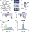
The roles of the basal ganglia (BG) in motor control are much debated. Many influential hypotheses have grown from studies in which output signals of the BG were not blocked, but pathologically disturbed. A weakness of that approach is that the resulting behavioral impairments reflect degraded function of the BG per se mixed together with secondary dysfunctions of BG-recipient brain areas. To overcome that limitation, several studies have focused on the main skeletomotor output region of the BG, the globus pallidus internus (GPi). Using single-cell recording and inactivation protocols these studies provide consistent support for two hypotheses: the BG modulates movement performance ('vigor') according to motivational factors (i.e. context-specific cost/reward functions) and the BG contributes to motor learning. Results from these studies also add to the problems that confront theories positing that the BG selects movement, inhibits unwanted motor responses, corrects errors on-line, or stores and produces well-learned motor skills.
Copyright © 2010 Elsevier Ltd. All rights reserved.
Figures


Similar articles
-
Testing basal ganglia motor functions through reversible inactivations in the posterior internal globus pallidus.J Neurophysiol. 2008 Mar;99(3):1057-76. doi: 10.1152/jn.01010.2007. Epub 2007 Dec 12. J Neurophysiol. 2008. PMID: 18077663 Free PMC article.
-
Shaping of motor responses by incentive values through the basal ganglia.J Neurosci. 2007 Jan 31;27(5):1176-83. doi: 10.1523/JNEUROSCI.3745-06.2007. J Neurosci. 2007. PMID: 17267573 Free PMC article.
-
The Avian Basal Ganglia Are a Source of Rapid Behavioral Variation That Enables Vocal Motor Exploration.J Neurosci. 2018 Nov 7;38(45):9635-9647. doi: 10.1523/JNEUROSCI.2915-17.2018. Epub 2018 Sep 24. J Neurosci. 2018. PMID: 30249800 Free PMC article.
-
Sensorimotor contributions of the basal ganglia: recent advances.Phys Ther. 1990 Dec;70(12):864-72. doi: 10.1093/ptj/70.12.864. Phys Ther. 1990. PMID: 2236229 Review.
-
A consideration of sensory factors involved in motor functions of the basal ganglia.Brain Res. 1985 Jun;356(2):133-46. doi: 10.1016/0165-0173(85)90010-4. Brain Res. 1985. PMID: 3924350 Review.
Cited by
-
Inter-Software Consistency on the Estimation of Subcortical Structure Volume in Different Age Groups.Hum Brain Mapp. 2024 Oct 15;45(15):e70055. doi: 10.1002/hbm.70055. Hum Brain Mapp. 2024. PMID: 39440570 Free PMC article.
-
Comparison of basal ganglia regions across murine brain atlases using metadata models and the Waxholm Space.Sci Data. 2024 Sep 27;11(1):1036. doi: 10.1038/s41597-024-03863-3. Sci Data. 2024. PMID: 39333155 Free PMC article.
-
Causal effect of COVID-19 on longitudinal volumetric changes in subcortical structures: A mendelian randomization study.Heliyon. 2024 Aug 30;10(17):e37193. doi: 10.1016/j.heliyon.2024.e37193. eCollection 2024 Sep 15. Heliyon. 2024. PMID: 39296245 Free PMC article.
-
Refinement of efficient encodings of movement in the dorsolateral striatum throughout learning.bioRxiv [Preprint]. 2024 Jun 6:2024.06.06.596654. doi: 10.1101/2024.06.06.596654. bioRxiv. 2024. PMID: 38895486 Free PMC article. Preprint.
-
Energy landscape analysis of brain network dynamics in Alzheimer's disease.Front Aging Neurosci. 2024 May 15;16:1375091. doi: 10.3389/fnagi.2024.1375091. eCollection 2024. Front Aging Neurosci. 2024. PMID: 38813531 Free PMC article.
References
-
- Mink J. The basal ganglia: focused selection and inhibition of competing motor programs. Prog Neurobiol. 1996;50:381–425. - PubMed
-
- Redgrave P, Prescott TJ, Gurney K. The basal ganglia: a vertebrate solution to the selection problem? Neuroscience. 1999;89:1009–1023. - PubMed
-
- Hikosaka O, Nakamura K, Nakahara H. Basal ganglia orient eyes to reward. J Neurophysiol. 2006;95:567–584. - PubMed
-
- Desmurget M, Turner RS. Testing Basal Ganglia motor functions through reversible inactivations in the posterior internal globus pallidus. J Neurophysiol. 2008;99:1057–1076. This study examines the contributions of the BG to motor control by performing in-depth analyses of the effects of GPi inactivation on visually-targeted reaching movements in non-human primates. The strongest and most consistent effects were slowing and hypometria for all directions of movement, which correlated with reductions in the magnitude of movement-related EMG. Contrary to several current hypotheses, the following aspect to motor control were preserved during GPi inactivations: (1) movement initiation; (2) on-line correction of misdirected reaches; and (3) sequencing of muscle activation including appropriate suppression of antagonist activity. - PMC - PubMed
-
- Schmidt L, d’Arc BF, Lafargue G, Galanaud D, Czernecki V, Grabli D, Schupbach M, Hartmann A, Levy R, Dubois B, et al. Disconnecting force from money: effects of basal ganglia damage on incentive motivation. Brain. 2008;131:1303–1310. This ground-breaking study provides detailed insight into the behavioral impairments associated with bilateral lesions of the BG in humans. Auto-activation deficit (AAD) has long been recognized in the clinical literature as a common correlate of damage to the BG. Here, Schmidt et al. found that AAD patients did not modulate grip forces in response to the monetary incentives offered in a task, even though the patients were able to modulate force levels appropriately in response to explicit instructions. Interestingly, galvanic skin responses suggested that the AAD patients retained affective awareness of the incentive value of different monetary rewards. The authors conclude that the BG links incentive motivation to the appropriate scaling of motor output. - PubMed
Publication types
MeSH terms
Grants and funding
LinkOut - more resources
Full Text Sources


