Normal age-related brain morphometric changes: nonuniformity across cortical thickness, surface area and gray matter volume?
- PMID: 20739099
- PMCID: PMC3026893
- DOI: 10.1016/j.neurobiolaging.2010.07.013
Normal age-related brain morphometric changes: nonuniformity across cortical thickness, surface area and gray matter volume?
Abstract
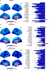
Normal aging is accompanied by global as well as regional structural changes. While these age-related changes in gray matter volume have been extensively studied, less has been done using newer morphological indexes, such as cortical thickness and surface area. To this end, we analyzed structural images of 216 healthy volunteers, ranging from 18 to 87 years of age, using a surface-based automated parcellation approach. Linear regressions of age revealed a concomitant global age-related reduction in cortical thickness, surface area and volume. Cortical thickness and volume collectively confirmed the vulnerability of the prefrontal cortex, whereas in other cortical regions, such as in the parietal cortex, thickness was the only measure sensitive to the pronounced age-related atrophy. No cortical regions showed more surface area reduction than the global average. The distinction between these morphological measures may provide valuable information to dissect age-related structural changes of the brain, with each of these indexes probably reflecting specific histological changes occurring during aging.
Published by Elsevier Inc.
Figures


Similar articles
-
Patterns of cortical degeneration in an elderly cohort with cerebral small vessel disease.Hum Brain Mapp. 2010 Dec;31(12):1983-92. doi: 10.1002/hbm.20994. Epub 2010 Mar 24. Hum Brain Mapp. 2010. PMID: 20336684 Free PMC article.
-
Age-related changes in the thickness of cortical zones in humans.Brain Topogr. 2011 Oct;24(3-4):279-91. doi: 10.1007/s10548-011-0198-6. Epub 2011 Aug 14. Brain Topogr. 2011. PMID: 21842406 Free PMC article.
-
Preservation of neuronal number despite age-related cortical brain atrophy in elderly subjects without Alzheimer disease.J Neuropathol Exp Neurol. 2008 Dec;67(12):1205-12. doi: 10.1097/NEN.0b013e31818fc72f. J Neuropathol Exp Neurol. 2008. PMID: 19018241 Free PMC article.
-
Healthy aging: an automatic analysis of global and regional morphological alterations of human brain.Acad Radiol. 2012 Jul;19(7):785-93. doi: 10.1016/j.acra.2012.03.006. Epub 2012 Apr 14. Acad Radiol. 2012. PMID: 22503890
-
Gray matter pathology in (chronic) MS: modern views on an early observation.J Neurol Sci. 2009 Jul 15;282(1-2):12-20. doi: 10.1016/j.jns.2009.01.018. Epub 2009 Feb 26. J Neurol Sci. 2009. PMID: 19249061 Review.
Cited by
-
Machine Learning-Driven Prediction of Brain Age for Alzheimer's Risk: APOE4 Genotype and Gender Effects.Bioengineering (Basel). 2024 Sep 20;11(9):943. doi: 10.3390/bioengineering11090943. Bioengineering (Basel). 2024. PMID: 39329685 Free PMC article.
-
Deep learning-based quantification of brain atrophy using 2D T1-weighted MRI for Alzheimer's disease classification.Front Aging Neurosci. 2024 Aug 14;16:1423515. doi: 10.3389/fnagi.2024.1423515. eCollection 2024. Front Aging Neurosci. 2024. PMID: 39206118 Free PMC article.
-
Relationship between domain-specific physical activity and cognitive function in older adults - findings from NHANES 2011-2014.Front Public Health. 2024 Jul 24;12:1390511. doi: 10.3389/fpubh.2024.1390511. eCollection 2024. Front Public Health. 2024. PMID: 39114526 Free PMC article.
-
Sex as a Determinant of Age-Related Changes in the Brain.Int J Mol Sci. 2024 Jun 28;25(13):7122. doi: 10.3390/ijms25137122. Int J Mol Sci. 2024. PMID: 39000227 Free PMC article. Review.
-
Cognitive reserve involves decision making and is associated with left parietal and hippocampal hypertrophy in neurodegeneration.Commun Biol. 2024 Jun 18;7(1):741. doi: 10.1038/s42003-024-06416-x. Commun Biol. 2024. PMID: 38890487 Free PMC article.
References
-
- Collins DL, Neelin P, Peters TM, Evans AC. Automatic 3D intersubject registration of MR volumetric data in standardized Talairach space. Journal of computer assisted tomography. 1994;18(2):192–205. - PubMed
-
- Courchesne E, Chisum HJ, Townsend J, Cowles A, Covington J, Egaas B, Harwood M, Hinds S, Press GA. Normal brain development and aging: quantitative analysis at in vivo MR imaging in healthy volunteers. Radiology. 2000;216(3):672–82. - PubMed
Publication types
MeSH terms
Grants and funding
LinkOut - more resources
Full Text Sources
Other Literature Sources
Medical


